Copyright 2010
Definition
Structure
The superior frontal gyrus is a narrow convolution between the supero-medial border of the hemisphere and the superior frontal sulcus. It has 2 segments: a dorsal and an orbitary. The dorsal segment is the one that borders the supero-medial border of the hemisphere; medially, it is limited by the cingulated sulcus. The orbitary segment, which is more inferior, occupies the inferior portion of the frontal lobe in the space between the interhemispheric fissure and the olfactory sulcus.
It has a diffuse layer IV, densely distributed pyramidal cells in the layer III, and larger ganglion cells densely distributed in layer V.
Function
It originates signals for volitional horizontal gaze saccades in the opposite eye field. It is also part of the signals that turn the head contralaterally.
The posterior part of the superior frontal gyrus acts as the supplementary motor area.
It is also thought to contribute to higher executive and cognitive functions (such as inductive reasoning, calculation) and to be responsible for working memory and sequence learning. It is also involved in the management of uncertainty and can anticipate pain and generate proper responses to attempt to avoid it.
Disease
The superior frontal gyrus is supplied by the anterior-medial division of the anterior cerebral artery. A stroke causing a lesion in this area may result in choreoathetotic and dystonic postures. Paralysis of contralateral eye turning and sometimes inability to turn the head may occur.
The superior frontal gyrus comprises one-third of the frontal lobe, bound by the superior frontal sulcus. It functions for self awareness, social response, and memory. Neuroimagining techniques including fMRI are used in diagnosis superior frontal gyrus dysfunction. Dysfunction in this area causes impairment in working memory and depression. Treatment of dysfunction is pharmocological for the allieviation of symptoms.

General View of the Gyri from the Lateral Surface of the Brain |
|
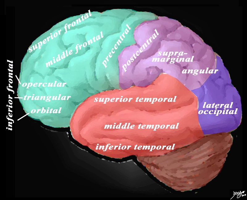
The lateral view of the brain shows the frontal lobe (green) parietal lobe (light mauve), the occipital lobe (purple) and the temporal lobe (red) In this view the frontal lobe gyri that are visible are; superior frontal, middle frontal, inferior frontal and precentral gyri. The inferior frontal gyri include the opercular, triangular and orbital. The parietal gyri include the post central, supramarginal, and angular gyri. The occipital gyrus that is visible is the lateral occipital. The temporal lobe gyri include the superior, middle and inferior temporal gyri. Courtesy Ashley Davidoff MD Copyright 2010 83029d05g.8 |

Superior Frontal Gyrus |
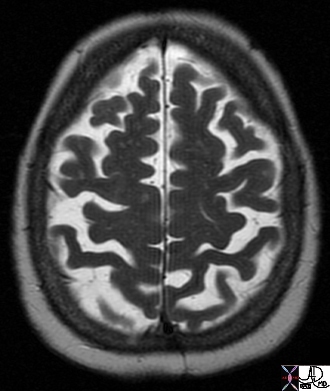
| 49051 brain central sulcus superior frontal gyrus Chronic Infarct Posterior Putamen normal T2 axial Courtesy Ashley Davidoff MD Uploaded RP |
