
The Common Vein Copyright 2010
Definition
Oligodendrocytes are cells of intermediate size, smaller than the astrocytes, and larger than microglial cells.
They have few, short processes, which remain in close relationship to the axons and neuronal cell bodies in the CNS, mostly present in the white matter. They have a dense cytoplasm rich in ribosomes and rough endoplasmic reticulum, large Golgi apparatus and abundant number of mitochondria. The processes of a single oligodendrocyte binds to the myelin sheath of numerous nerve fibers.
The oligodendroglia play an important role in the formation and maintenance of the myelin sheath of nerve fibers in the central nervous system; in peripheral nerve fibers, this function is assured by Schwann cells. Myelin is extremely important, since it increases impulse speed.
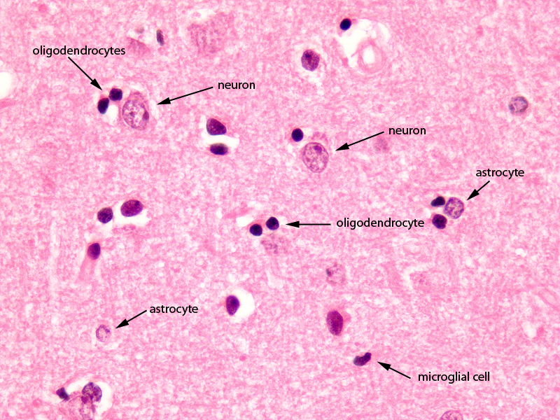
Cerebral Cortex Exemplifying the Oligodendrocyte Magnification 60X |
|
The image shows a histological section of the brain at 60X magnification. The section shows the 4 of the common basic types of brain cells; the largest is the neuron which is only found in gray matter. They are 10 times larger than the other cells but less common than the second type of cell called the glial cell. The astrocyte is a type of glial cell and is the second largest cell. It often lies in close association with the blood vessels. The third type of cell seen in this section, also a glial cell is the oligodendrocyte. It is smaller than the astrocyte and has a darker nucleus. It assists in the myelination of the white matter. It is in fact the predominant cell of white matter. The last cell seen in this image is the microglial cell. It is the smallest and has a fusiform shape. Courtesy of Thomas W.Smith, MD; Department of Pathology, University of Massachusetts Medical School. 97803 |
Injury to oligodendrocytes result from demyelinating diseases and leukodystrophies. Damage to developing oligodendrocytes in areas around the cerebral ventricles result in cerebral palsy.
The human brain contains roughly 84.6 billion glia and 86.1 billion neurons. (Azevedo)
Most cerebral cortex glia are oligodendrocytes (75.6%) then astrocytes (17.3%) and least for microglia (6.5%) (Pelvig)
The amount of glial celss that are found in brain tissue is related to brain size
| animal
|
% glial cells
|
| nematode | few glia |
| fruitfly | 25% |
| mouse | 65% |
| human | 90% |
| elephant | 97% |
(Allen)
References
Allen NJ, Barres BA. (2009). Neuroscience: Glia – more than just brain glue. Nature. 457(7230):675-7. PMID 19194443
Azevedo FA, Carvalho LR, Grinberg LT, Farfel JM, Ferretti RE, Leite RE, Jacob Filho W, Lent R, Herculano-Houzel S. (2009). Equal numbers of neuronal and nonneuronal cells make the human brain an isometrically scaled-up primate brain. J Comp Neurol. 513(5):532-41. PubMed
Pelvig DP, Pakkenberg H, Stark AK, Pakkenberg B. (2008). Neocortical glial cell numbers in human brains. Neurobiol Aging. 29(11):1754-62.(figures given are those for females) PubMed