Transverse or Axial Projection – Concepts
The Common Vein Copyright 2010
Introduction
The Bacward and Downward Facing ‘C’s” of the Brain in the Axial Plane Ventricular System (light blue) in Central Position |
|
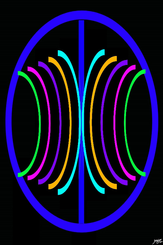
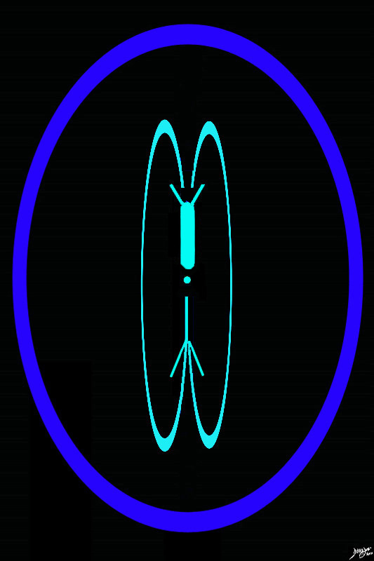
The anchoring concept of the brain in the axial plane is two cerebral hemispheres with a series of structures reminiscent of backward and downward facing “C’s” symettrically positioned around the center. 93914.3ka09.8sd01 Courtesy Ashley DAvidoff MD Copyright 2010 all rights reserved |
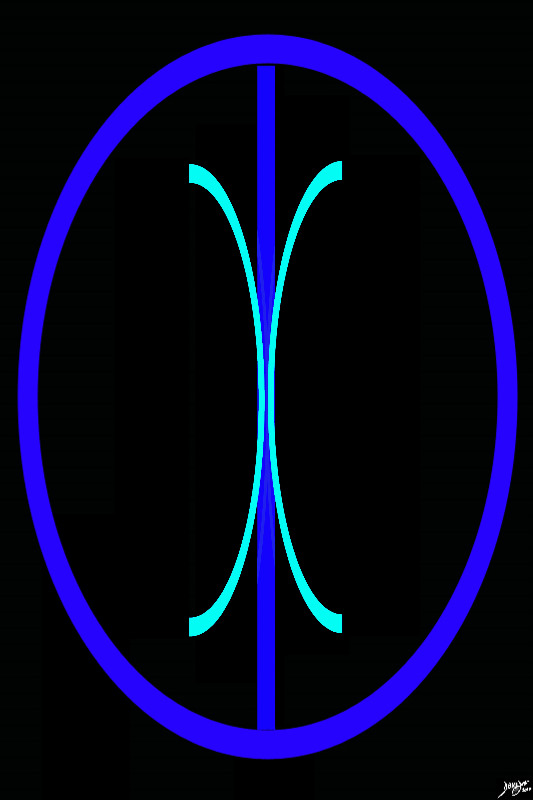
The Innermost Layer – The Ventricular System |
|
The most central, backward and downpointing ‘C” is the ventricular system (teal) Courtesy Ashley Davidoff MD copyright 2010 93914.3ka03d01.8s |
|
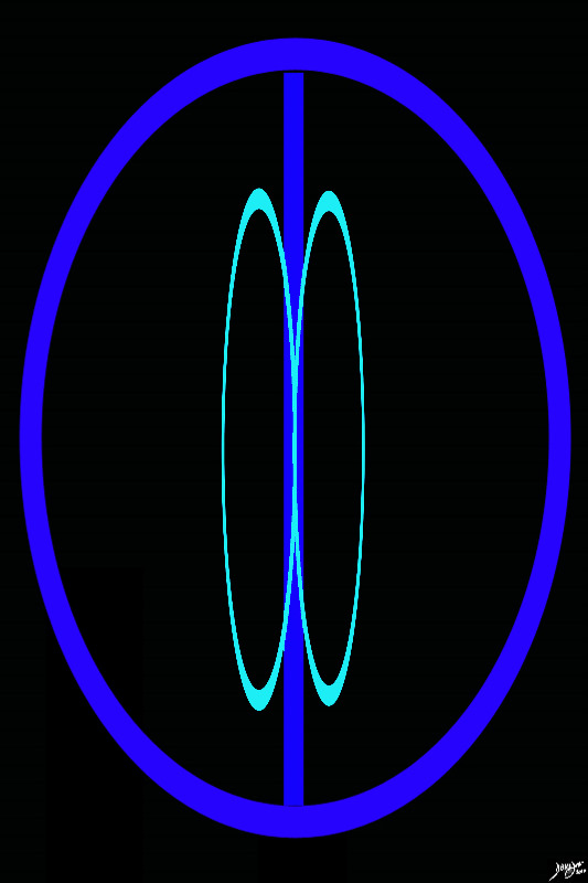
The Transverse Plane |
|
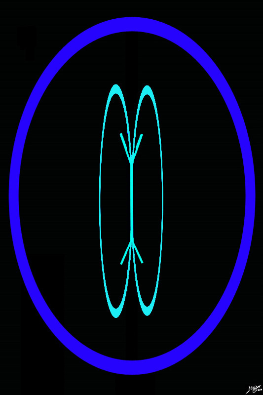
The axis of the brain in the sagittal view consists of an anteroposterior or relatively horozontal vector and a relatively vertical, craniocaudal vector. The ventricular system folows these vectors and consists of a paired horizontal system and a single vertical system Courtesy Ashley Davidoff MD copyright 2010 all rights reserved 94458d02.8s |
The Lateral Ventricles |
|
The lateral ventricles are the first components of the ventricular system to appear as the planes advance from the top of the brain caudally. They are almost H shaped as they start to appear and retain a semblance of this shape for a number of cuts. Courtesy Ashley Davidoff copyright 2010 all rights reserved 93914.3kd03b02b01.81sd02 |
Form – Two Anterior Horns and Two Posterior Horns |
|
The anterior and posterior aspect of the lateral ventricle diverge from the body to form structures reminiscent of horns and hence the name anterior or frontal horns and posterior or occipital horns. Courtesy Ashley Davidoff copyright 2010 all rights reserved 93914.3kd03b02b01.81sd02 |
|
The Lateral Ventricles |
|
This T2 weighted image is taken in the transverse or axial plane when the superior aspect of the lateral ventricles are first clearly seen as the scan advances in the forebrain region from superior to inferior. The H shape is characteristic at this level, and the ventricles are most voluminous at this level in the axial plane Courtesy Ashley Davidoff MD copyright 2010 all rights reserved 94077c01.81s |
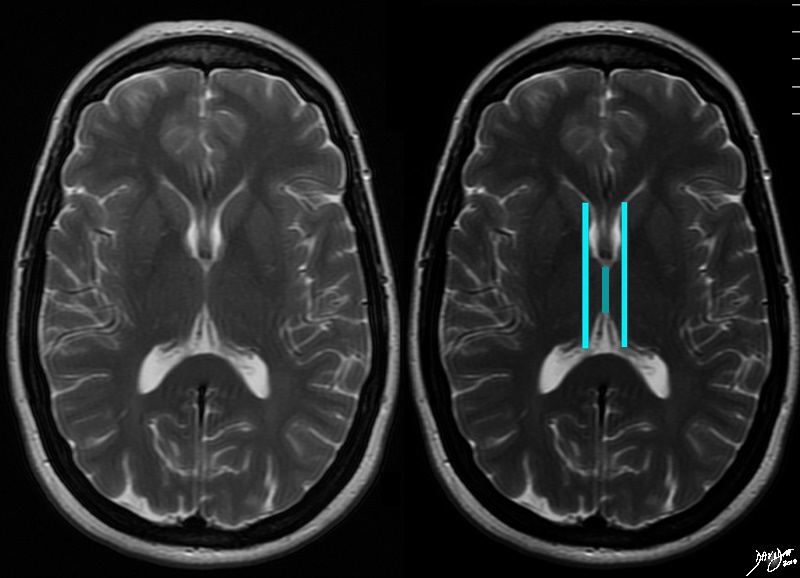
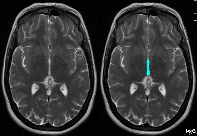
The Third Ventricle |
|
The 3rd ventricle in the axial projection is shown as a slit like structure posterior to the frontal horns. It is very narrow heing longer in the A-P dimension than it is in the transverse direction creating a a shape reminiscent of a sausage. It is deceptively large in its craniocaudad span. It is seen in a variety of shapes including a narrow oblong, narrow diamond, and sometimes even as keyhole shaped structure. Courtesy Ashley Davidoff copyright 2010 all rights reserved 93914.3kd04b.82sd02 |
3rd Ventricle |
|
The 3rd ventricle in the axial projection is very narroe and is linger than it is narrow, but is deceptively large in its craniocaudad span. Its shape varies from a narrow oblong, to a narrow diamond, to a keyhole shaped structure. Courtesy Ashley Davidoff MD copyright 2010 all rights reserved 94080c01.81s |
Aqueduct of Sylvius – Cerebral Aqueduct |
|
The aqueduct of Sylvius is the next structure to come into focus as we proceed inferiorly It is a tiny rounded structure that is located in the posterior aspect of the midbrain.It is barely visible on most imaging studies and it lies posterior and inferior to the third ventricle. Courtesy Ashley Davidoff copyright 2010 all rights reserved 93914.3kd05b.84sd02 |
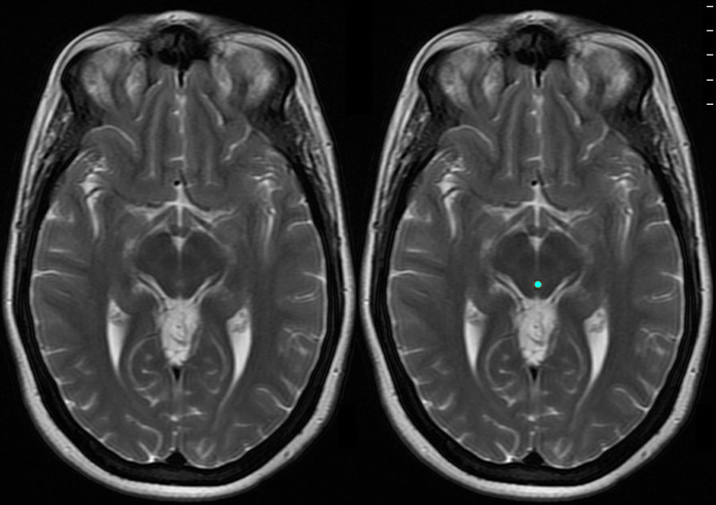
Aqueduct of Sylvius Cerebral Aqueduct in the Midbrain |
|
The axial T2 weighted MRI image through the midbrain shows the small but all important aqueduct of Sylvius (cerebral aqueduct) as it passes through the midbrain (Mickey Mouse appearance). Courtesy Ashley Davidoff MD copyright 2010 all rights reserved 94081c01.81s |

The 4th Ventricle |
|
The 4th ventricle is the last landmark of the ventricular system seen as we proceed inferiorly. It is seen as a puffed crown or the pontifs hat (mitre), which lies posterior to the pons (ponntiff /pons) inferior to the aqueduct of Sylvius and anterior to the cerebellum. It lies within the hinbrain. Courtesy Ashley Davidoff copyright 2010 all rights reserved 93914.3kd06bd03.8s |
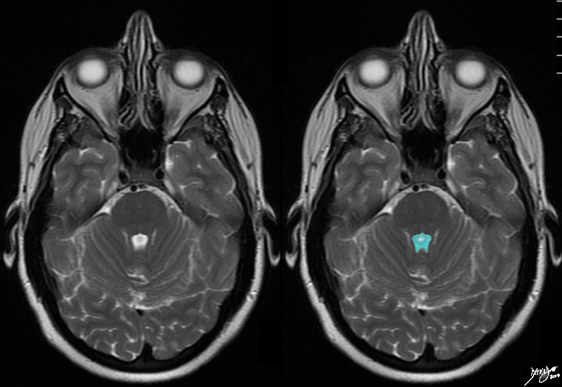
The 4th Ventricle |
|
The T2 weighted axial image through the 4th ventricle is chatracterised by the “puffed crown” or pontiffs hat of the pons of this ventricle. Identifying the 4th ventricle allows localisation of the cut to the hindbrain. The 4th ventricle is anterior to the cerebellum and posterior to the pons. Courtesy Ashley Davidoff MD copyright 2010 all rights reserved 94084cd05.81s |
|
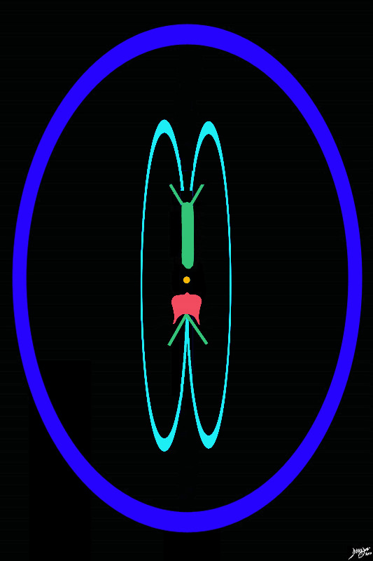
The Distribution of the ventricles in the Brain Forebrain (green) Midbrain (orange) and hindbrain (salmon) |
|
This diagram shows the positioning of each of the components of the ventriclar system . The lateral ventricles and third ventricle lie in the forebrain (green), the aqueduct of Sylvius (orange) lies in the midbrain, and the 4th ventricle (salmon) lies in the hinbrain. Courtesy Ashley Davidoff copyright 2010 all rights reserved 93914.3kd06bd04.8s |
|
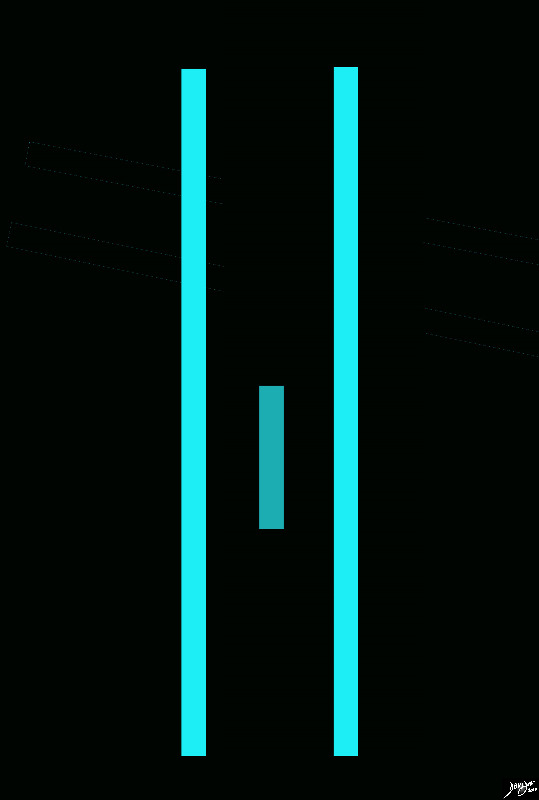
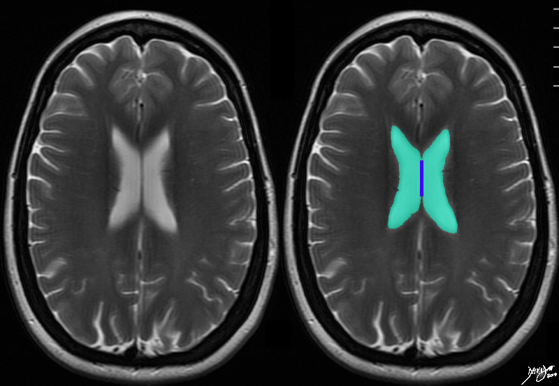
Axial T2 weighted Image |
|
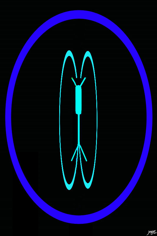
The horizontal limbsreviewed in the sagittal section are reflected as two parallelvectors that include parrt of the frontal horns and occipital components, while the teal green limb in the middle represents the conglomerate vertical limb that includes the third ventricle, the aqueduct of Sylvius and the fourth ventricle. Courtesy Ashley Davidoff MD copyright 2010 all rights reserved 94458e08a.8s |