
The Common Vein Copyrighrt 2010
Definition
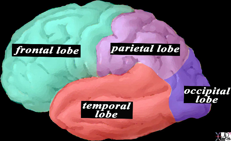
The parietal lobe is part of the forebrain and lies between the frontal lobe abnd occipital lobe. It plays an important role in integrating information from different sensory modalities (vision, touch, hearing). It is particularly involved in the perception of space and attention and, more specifically, the superior parietal cortex is involved in the visuomotor control of movements, including saccades eye movements.
Structure The parietal lobe occupies the middle and superior part of the hemisphere, being limited posteriorly by the parieto-occipital sulcus, anteriorly by the central fissure and below by the Sylvian fissure. It contains a fissure (the intraparietal sulcus) that further splits it into postcentral and horizontal sulci that separate 3 convolutions (posterior central, supramarginal and angular gyri).
Function The parietal lobes contain the postcentral gyrus, which is essentially a topical representation of the somatic sensory innvervation to various contralateral parts of the body with the more sensitive areas receiving more representation (sensory homunculus).
Caudal to this area, the parietal lobe is divided into a superior and inferior lobe. The inferior portion is responsible integration of information for speech. The superior portion is important for the recognition of the surrounding environment, including oneself.
The parietal lobes of the cerebral cortex are able to organize information from the senses regarding the shape, texture and weight and transform them into general perceptions. Mathematical skills and language have their origin somewhere in that area, more specifically in the areas adjacent to the temporal lobes. The parietal lobes also help people to orient themselves in space and realize the position of body parts.
Disease A deficit in the anterior part of the parietal lobes produces numbness on the opposite side of the body. Individuals with larger lesions may develop apraxia and awareness of the right-left directions. The ability to recognize parts of the body or the space can become impaired, sometimes causing the individual to ignore the seriousness of their problem and neglect or even deny the existence of paralysis affecting one side of the body opposite the brain lesion. Gerstmann’s syndrome, Balint’s syndrome, and hemispheral neglect are all associated with parietal lobe dysfunction.
Treatment would involve therapy in less extreme cases, and permanent care assistance for those with extreme damage.
Context
The Parietal Lobe (lime green) Part of the Forebrain (Prosencephalon) Member of the Cerebrum (Telencephalon) Member of the Cerebral Hemispheres |
|
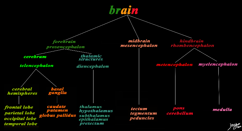
The basic and simplest classification of the brain into forebrain midbrain and hindbrain is shown in this diagram and advanced to a more complex tree using the embryological and evolutionary terminologies. The forebrain consists of the cerebrum also called the prosencephalon, which contains the more advanced form of the brain and the thalamic structures which contain more basic structures. The cerebrum (telencephalon) itself consists of two cerebral hemispheres and paired basal ganglial structures. Each cerebral hemisphere will have gray and white matter distributed in the frontal parietal temporal and occipital lobe, with the basal ganglia being part of the gray matter deep in the cerebral hemispheres. The most important thalamic structures arising from the diencephalons include the thalamus itself and the hypothalamus. The midbrain (mesencepaholon) consists of the tectum tegmentum and cerebral peduncles. The hindbrain has two major branch points based on the evolutionary development. The pons and cerebellum(part of the metencephalon) are grouped and the medulla (part of the myelencephalon is the second branch. Courtesy Ashley Davidoff MD Copyright 2010 All rights reserved 97686.8s |
|
Conceptual Framework of the Brain |
|
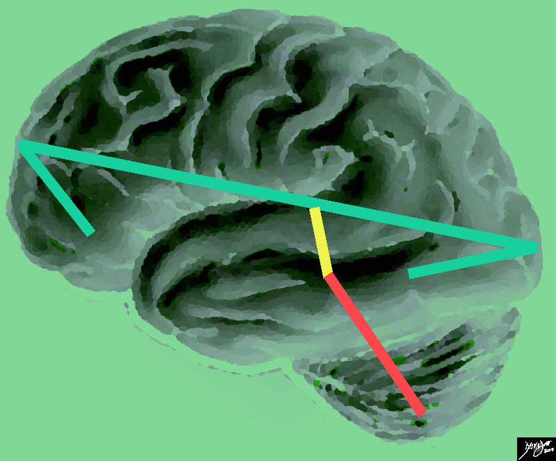
The vector of the forebrain (green) is folded at its anterior aspect as a projection of the frontal lobe and the posterior fold forms the temporal lobe. The midbrain (yellow has its axis just tipped slightly anteriorly and the hindbrain (salmon red) is tipped more posteriorly Courtesy Ashley Davidoff MD copyright 2010 all rights reserved |
The Central Sulcus is the Border between the Frontal Lobe and the Parietal Lobe |
|
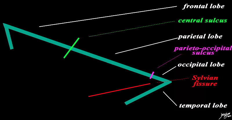
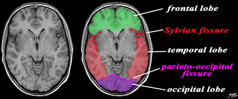
The forebrain is divided into the frontal lobe anetriorly, separated by the central sulcus (bright green), the parietal lobe separated from the occipital lobe by the parieto-occipital fissure (pink), and the temporal lobe separated from the frontal and occip[ital lobe by the Sylvian fissure (lateral sulcus) (red). The distinction between the occipital and temporal lobe is more of a functional division than a structural division Courtesy Ashley Davidoff MD copyright 2010 93887b03b05.8s |
|
MRI Showing the Central Sulcus and the Lobes |
|
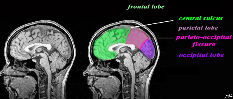
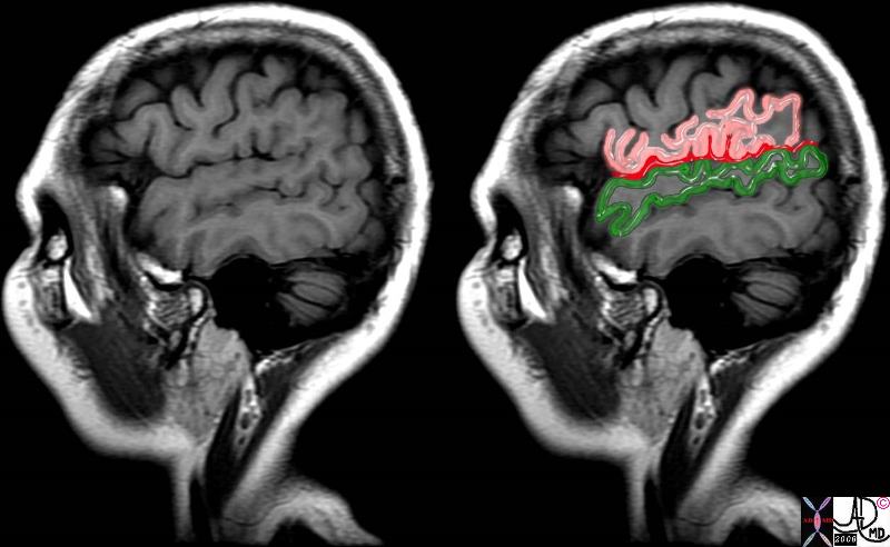
The sagittal image is deeper in the brain near the midline through the interhemispheric fissure, and is intended to demonstrate the the parieto-occipital fissure (pink) in order to define the border between the parietall and occipital lobe. The central sulcus (pink) has also been inferred in to demonstrate the border between the frontal lobe and parietal lobe. It is not usually seen in this projection. The Sylvian is also not usually seen in this projection. What we do start to appreciate are the layers of inverted c shaped structures. MRI 3D FFE Courtesy Philips Medical systems rendered by Davidoff art 92141c01label.82s |
|
83029d13.8s |
|
This is a diagram looking at the forebrain from the side showing the anteriorly placed frontal lobe, inferiorly placed temporal lobe posteriorly placed occipital lobe and superiorly placed occipital lobe. In this view the size of the frontal lobe dominates and in fact it is the largest portion of the forebrain. Courtesy Ashley DAvidoff MD copyright 2010 all rights reserved 83029d13.8s |
Somatosensory Region Parietal Lobe Above the Sylvian Fissure (red) and Temporal Lobe Below |
|
The sagittal T1 weighted MRI on the lateral aspect of the brain shows the Sylvian fissure (red) above which is the parietal lobe, and below which is the temporal lobe. 71060c07.8s brain pain pathway somotosensory cortex S 2 SII operculum parietal lobe temporal lobe frontal lobe lateral sulcus Sylvian fissure MRI T1 weighted Courtesy Ashley Davidoff MD copyright 2008 |
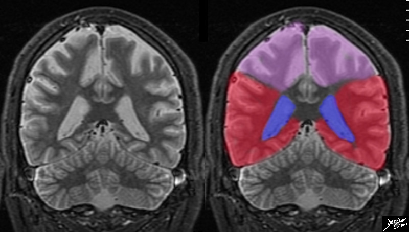
Junction of the Parietal Lobe (pink) and Temporal Lobe (green) at the Operculum |
|
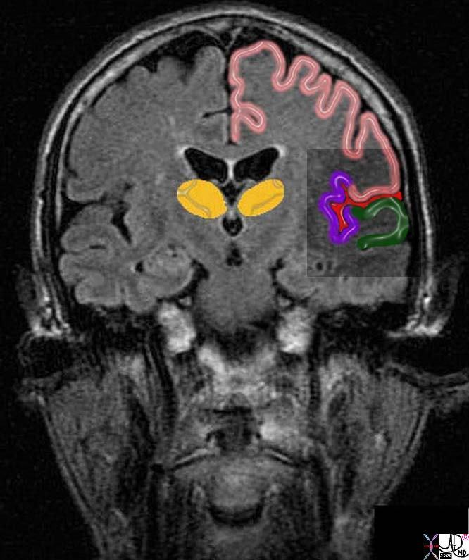
The coronal T1 weighted image of the brain reveals the deeper components and the junction of the insular cortex and its neighbours. The lateral somatosensory cortex is centered around the Sylvian fissure (red) It incorporates the insula (purple) the upper lid of the operculum which is part of the parietal cortex (pink) as well as parts of the frontal cortex, and the lower lid of the operculum (green) which is part of the temporal lobe. Courtesy Ashley Davidoff MD copyright 2010 38610c06b07.8s |
|
Axial Framework of the Forebrain |
|
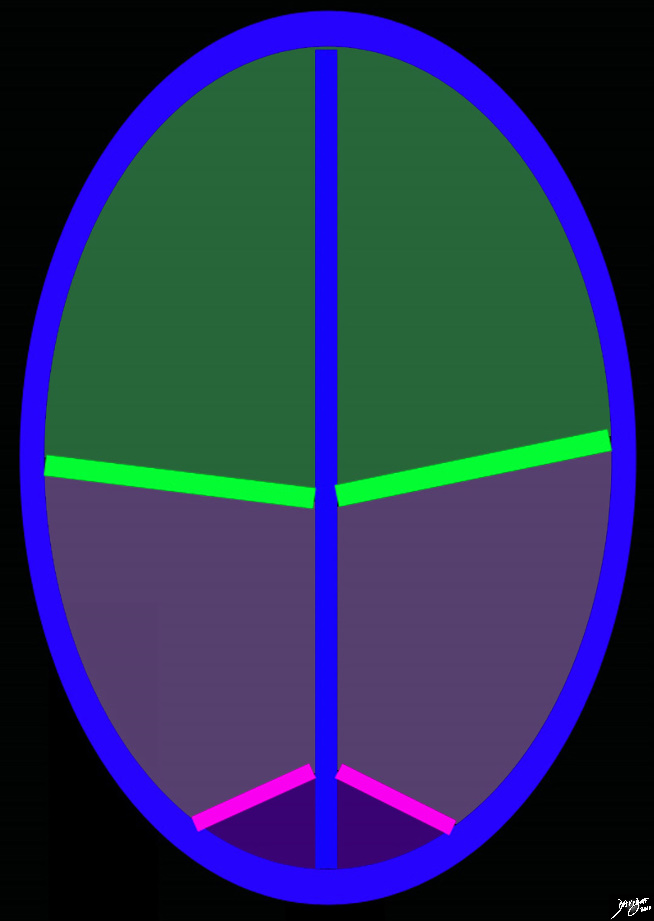
The diagram reflects the interhemispheric fissure as depicted in the axial plane dividing the brain into two halves. The bright green line represents the central sulcus which separates the the frontal lobes (green) from the parietal lobes (light purple. The parito-occipital fissure (pink) separates the parietal lobes from the occipital lobes. By convention anterior is on the top and posterior on the bottom, while the patients right is to our left and vice versa for the patients left Copyright 2010 Courtesy Ashley Davidoff MD 93914.85sb |

The Central Sulcus Frontal Lobe and Parietal Lobe in the Transverse Plane |
|
The diagram of the brain as viewed from above shows the interhemispheric fissure, and the central sulcus (lime green) which divides tyhe brain into the frontal lobe anteriorly and the parietal lobe posteriorly. The presentral gyrus, (blue) is the motor gyrus and is a landmark posterior border of the frontal lobe. The post central gyrus (pink) is the sensory gyrus and is the landmark anterior border of the parietal lobe. Courtesy Ashley Davidoff MD Copyright 2010 All rights reserved 52981b05.8.44kb01c06.9c |
|
Axial Projection MRI |
|
The axial image is intended to demonstrate ttwo important fissures; the Sylvian fissure (in red) the parieto-occipital fissure (pink) in order to demonstrate the border between the frontal and temporal lobe in the axial plane and the junction of the temporal and occipital lobe MRI SENSE Courtesy Philips medicaL systems rendred by Davidoff art 92142c06b01 label.8s |
The Sylvian Fissure, Parietal Lobe, and Temporal Lobe |
|
In this T2 weighted MRI image – the forebrain (green is seen centered aroubnd the ventriclar system. The red linees indicate the Sylvian fissures which in this instance in a relatively posterior cut reflect the parietal lobe superiorly and the temporal lobes inferiorly (darker green) The temporal lobes rest of the tentorium (white curvilinear convex lines). below the tenntorium is the midbrain and hind brain Courtesy Ashley Davidoff MD copyright 2010 all rights reserved 89721c02b03.8s 89721c02b03 |
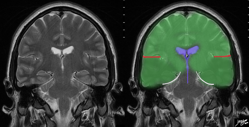
Posterior Coronal View Parietal and Temporal Lobes |
|
The coronal MRI shows the diversion of the ventriclesand their course inferiorly into the temporal horns allowing us to place the cut fairly posteriorly, and beyond the central sulcus, thus the superior aspect of the cut represents the parietal lobes, and the forebrain lying above the tentorium must represent the temporal lobes (red). Courtesy Ashley Davidoff MD copyright 2010 all rights reserved 72246c02.8s |
Applied Anatomy
Ischemia and Infarction of the Parietal Lobe
Acute Infarctions
Acute Infarction Right Parietal Lobe |
|
The diffusion weighted MRI in axial projection shows a high intensity region in the right parietal and right temporal region revealing an acute infarction poattern in these regions reflecting right middle cerebral artery territory. Note that the basal ganglia have been spared, but the insula and operculum are involved. Courtesy Ashley Davidoff MD copyright 2010 71275c01c03 |
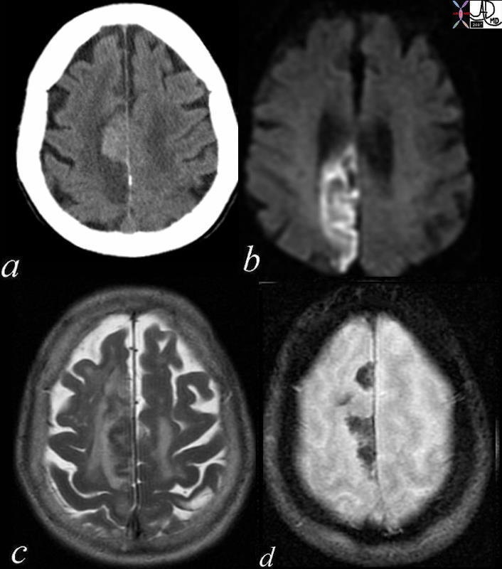
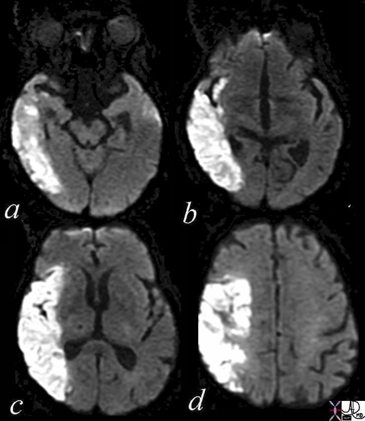
Acute Embolic Infarct High Parietal Region |
|
The axial images are from a patient with atrial fibrillation and neurological deficits. Image a is a CT scan which shows a high density lesion i the vertex of the right pariettal lobe suggesting hemorhagic change. Image b is a diffusion weighted MRI image at the level of the ventricles which shows a high intensity region in the parieto-occipital region suggesting acute infarction. Image c is a axial T2 weighted image showing edema in the white matter of the right parietal lobe. Image is aGRE image showing mixed heterogeneity with probable iron deposition suggesting subacute or chronic hemorhage. Findings are consistent with old and new multicentric infarcts of the brain likely from the heart caused by atrial fibrillation Courtesy Ashley Davidoff MD copyright 2010 71239c01 |
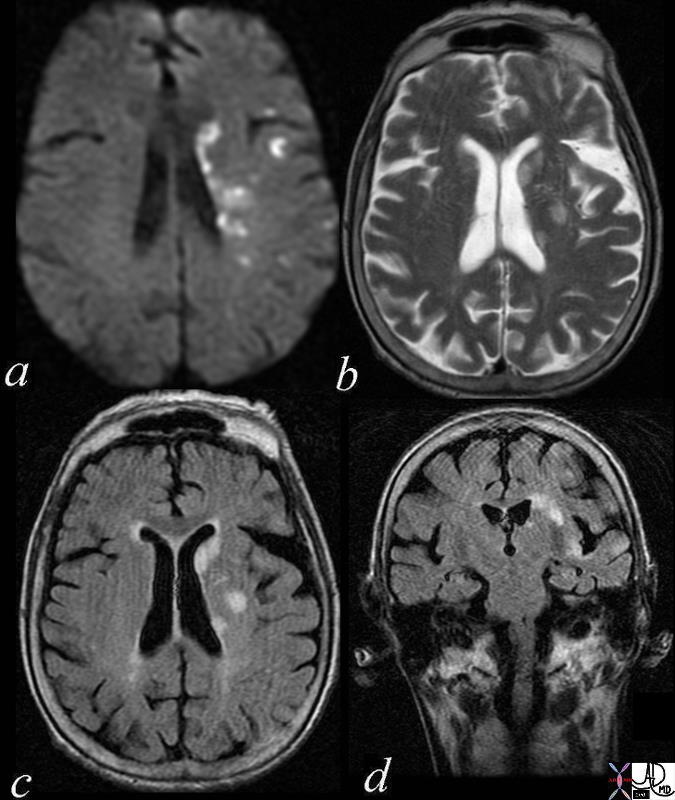
Multicentric Infarcts – Carotid Stenosis |
|
The images reveal multicentric acute infarcts in the left cerebral hemisphere involving the internal capsule and left parietal lobe cortex. Image a is the DWI sequence and the high intensity foci are diagnostic of acute infarction. Image b is a T2 weighted image that reflects increase water in the high intensity regions. Image c is an axial FLAIR and d a coronal FLAIR sequence both sensitive to the regions of infarct and characerized by high intensity foci in the regions of acute infarction. brain cerebral multicentric infarcts internal capsule parietal lobe cortex dx multicentric infarct left cerebral hemisphere associated with a left carotid stenosis with presumed showering of the embolic material embolism a = DWI b = T2 weighted imaging c – FLAIR axial d FLAIR coonal ask MRI Davidoff MD 72014c01 |
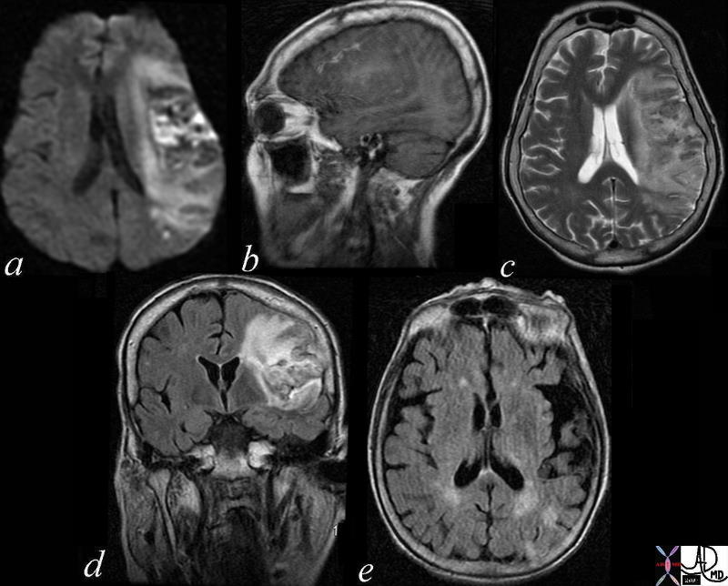
Subacute Hemorrhagic Infarct of the Parietal Lobe MRI |
|
The series of MRI images from a 70 year old male who by history suffered a stroke 1 month ago and has new onset symptoms. the series of images reveal complex changes of a subacute hemorrhagic infarct. Image a is from a DWI sequence and it shows a heterogenous increase in signal some of which represents T2 shine throughand sopme of which is bright raising the question on an acute on subacute entity. Image b is a sagittal T1 weighted image which shows areas of vague increase in density suggesting hemorrhage. Image c is a T2weighted sequence and shows some increase in water but the granular low intensity suggests hemosiderin deposit. Image d is FLAIR sequence showing increase brightness to the lesion in the left parietal lobe and image e is an axial FLAIR sequence The findings suggest extensive infarct in the left MCA territory which has mild mass effect on ventricles with petechial hemorrhage as seen on T2 and FLAIR and hyperintense T2 shine through on diffusion weighted images The punctate areas in left parietal lobe with restricted area of diffusion raises the question of a recent small infarct less bright regions on DWI suggests a subacute hemorrhagic infarct. Courtesy Ashley Davidoff MD copyright 2010 71000c03 |
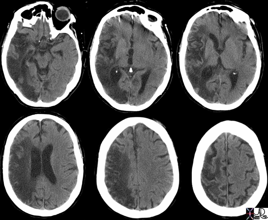
Chronic Parietal Infarction – CT scan |
|
The CT scan shows the effects of an extensive right middle cerebral infarct and involves the right parietal lobe, temporal lobe, and occipital lobe. The right lateral ventricle is enlarged because of the loss of brain tissue as a result of the infarction. This process is called encephalomalacia (brain softening) and the changes are called ex vacuo changes because the loss of tissue results in gain of space resulting in the shift of the ventricle into the space. Courtesy Ashley Davidoff MD copyright 2010 46031c |
Chronic Parietal Infarction with Encephalomalacia |
|
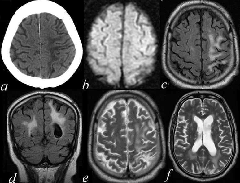
The series of axial and coronal images are from a 74 year old man with mental status changes. Image a is an axial CT scan showing a region of white matter hypodensity over the high parietal region. Image b is a DWI image that shows no abnormality in the region thus excluding an acute infarct. Image c is a FLAIR sequence in the axial plane and image d is a FLAIR seqience in the coronal plane suggesting increase water in the white matter of the high left parietal region confirmed by the T2 weighted sequence of e. The last axial image is a T2 weighted sequence through the ventricles showing that the left lateral ventricle is larger than the right. These findings suggest a chronic left MCA infarct with gliosis, encephalomalacia and ex vacuo changes. Courtesy Ashley Davidoff MD 71411c01.801 |
Metastatsis Parietal Lobe |
|
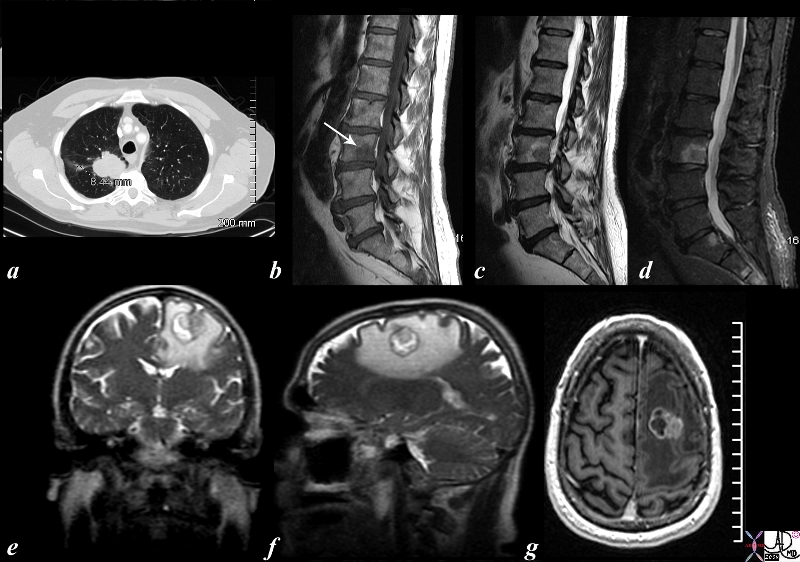
The CTscan and MRI images are from a 56 year old male who presented with back pain and a neurological deficit. The CTs scan of the chest shows a right upper lobe lung mass and the MRI of the spine (b= T1, c =T2 and d= STIR) confirm the presence of a metastasis in L3) while MRI of the brain (e= T2 coronal, f = T2 sagittal and g= post gadolinium study) reveal a mass in the high left parietal lobe with surrounding edema (white around the round tumor in e and f) and showing enhancement (g). Courtesy Ashley Davidoff MD copyright 2010 87681c02.8s |