
Copyright 2010
Introduction
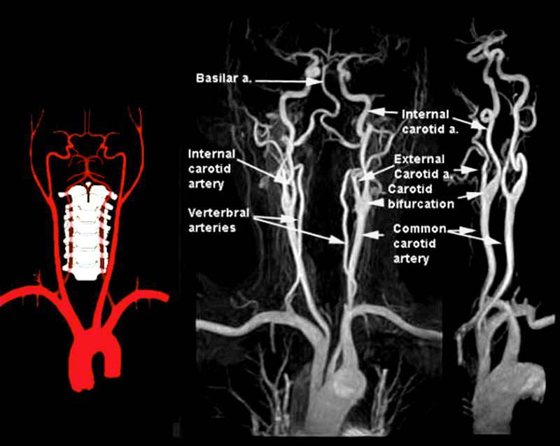
The right and left vertebral arteries arise from the right and left subclavian arteries at the base of the neck. From C6, they flow vertically in the transverse channel dug in the transverse processes of cervical vertebrae. After circling the lateral mass of the atlas, each vertebral artery perforates the dura and the foramen magnum. They head to the anterior surface of the medulla oblongata to the bulbo-pontine sulcus. Each artery fuses here with the opposite counterpart to form the basilar artery.
Overview |
|
The diagram shows the main branches of the blood supply to the brain which includes the carotid and vertebro-basilar systems. The carotid system supplies the brain from the internal carotid – a branch of the common carotid which arises from the aorta. On the right side the common carotid arises from the brachiocephalic artery and on the left it arises as the common carotid as the second branch off the aortic arch It enters the cranial cavity through the cavernous sinus and gives rise to two main branches; the anterior cerebral and the middle cerebral The vertebral arteries usually arise from the subclavian arteries and then travel in the vertebral foramina in the cervical spine. The basilar artery is formed by the two vertebral arteries and travel as a single artery over the upper medulla and the entire pons. Its terminal division is into the right and left posterior cerebral arteries. Its first branch after this division is the posterior communicating artery. It continues around the pons and midbrain in the ambient and quadrigeminal cisterns and then ascends above the tentorium to end in the occipital lobe. Each of the vessels contributes to the circle of Willis through communicating arteries. The vertebro-basilar system provides the posterior communicating arteries bilaterally and the carotid system provides the anterior communicating arteries via the middle cerebral artery. The PICA or posterior inferior cerebellar artery arises from the vertebral arteries and the anterior inferior cerebellar artery arises from the basilar artery. The superior cerebellar artery arises from the tip of the basilar artery just before it terminates in the posterior cerebral arteries Courtesy Philips Medical Systems 92492.8 |
Basic Components of the Vertebrobasilar Sytem |
|
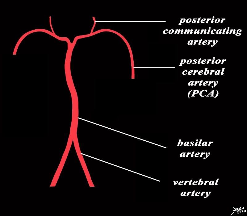
The basilar artery is formed by the two vertebral arteries and travels as a single artery over the upper medulla and the pons It gives rise to the posterior cerebral artery (PCA) and the first branches off the PCA are the two posterior communicating arteries that feed into the circle of Willis Image Courtesy Ashley Davidoff MD Copyright 2010 All rights reserved 97194b06b.82s |
Vascular Distribution |
|
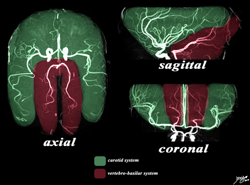
There are two systems that supply the brain with blood. The MRA depicted in the axial sagittal and coronal views depict the general areas covered by the two systems. The carotid system gives rise to the anterior and middle cerebral arteries which cover the green area. The vertebro-basilar system, mostly through the posterior cerebral artery and cerebellar arteries, supply the maroon portion. Image Courtesy Philips Medical Systems Artistic rendering Davidoff MD Copyright 2010 92479c03.8s |
Concepts – Feeding into the Circle of Willis |
|
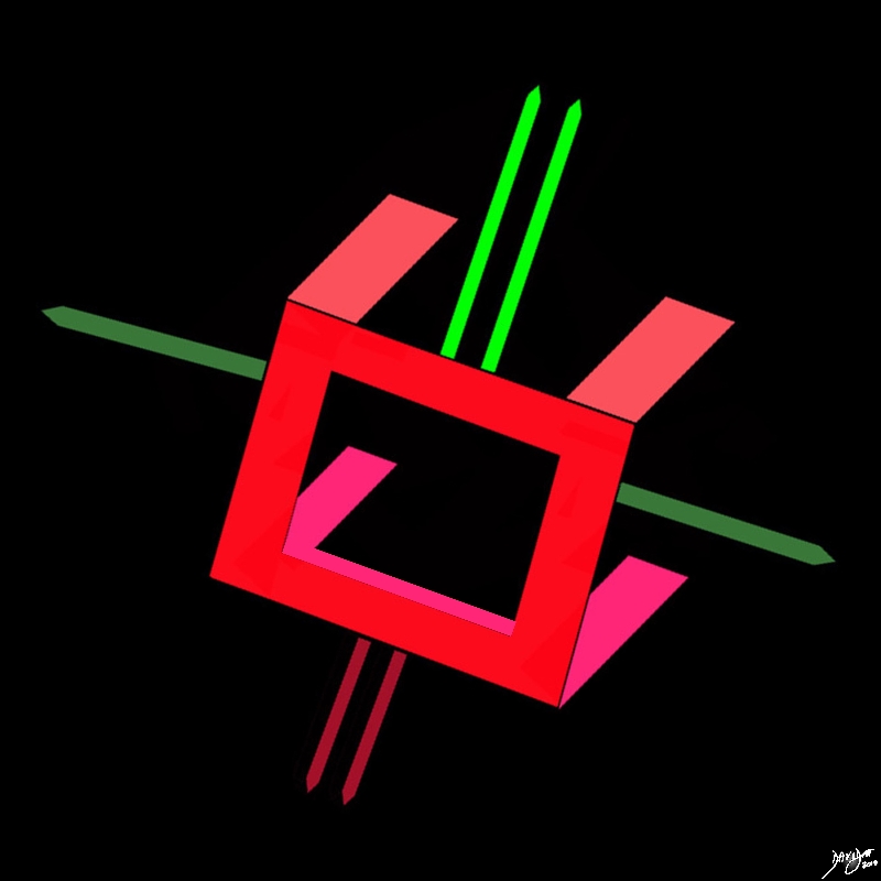
The cerebral circulation has a unique system that allows for compensatory flow when any one of the 4 vessels is narrowed or occluded. This system is centered on the circle of Willis. The brain has two basic systems that feed it; the carotid system (in this case salmon pink) and the vertebro-basilar system (brighter pink). They both feed into the circle of Willis (bright red) via communicating branches. The middle cerebral artery is the vessel that feeds into the COW by providing the anterior communicating artery and the posterior cerebral artery that feeds the COW by providing the posterior communicating artery. Conceptually as depicted in this diagram the circle of Willis is the centre of the cerebral circulation and from it blood flows into the anterior (bright green), middle (darker green) and posterior cerebral (maroon) regions. Image Courtesy Ashley Davidoff MD Copyright 2010 97194b16bgvert02.8s |
Contributions from Each of the Circulations |
|
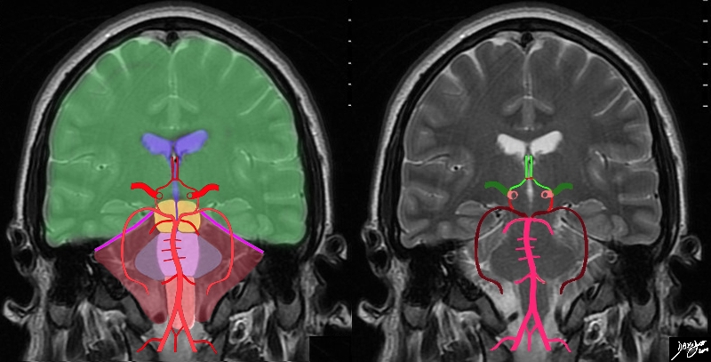
The diagram shows the main branches of the blood supply to the brain including the circle of Willis overlaid on coronal MRI image to portray the approximate position of the vessels in the brain. The image on the right shows the combined system all in red, and the image on the right shows the derivation from the vertebrobasilar and carotid systems The carotid system supplies the brain from the internal carotid (salmon pink). We demonstrate its terminal bifurcation into middle cerebral (dark green) and anterior cerebral (bright green). The anterior communicating artery runs between the two anterior cerebrals (bright red) The basilar artery (pink) is formed by the two vertebral arteries and it travels as a single artery over the upper medulla and the pons. Its terminal branch is the posterior cerebral artery (maroon). The first branch off the posterior cerebrals is the posterior communicating which joins the middle cerebral to complete the circle of Willis Each of the carotid and vertebro-basilar systems contributes to the circle of Willis through communicating arteries. The vertebro-basilar system provides the posterior communicating arteries bilaterally from the posterior cerebral and the carotid system provides the anterior communicating arteries via the anterior cerebral arteries. Courtesy Ashley Davidoff MD Copyright 2010 All rights reserved 89721c06b.8sg05.8s |
Major Branches of the Vertebro-basilar System |
|
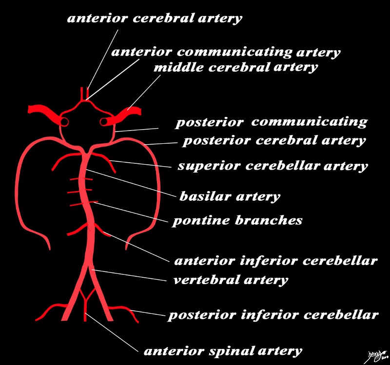
The diagram shows the main branches of the vertebro-basilar system. As the vertebral arteries emerge through the foramen magnum they give rise to the posterior inferior cerebellar artery. Shortly thereafter and just prior to converging to form the basilar artery, each of the vertebral arteries gives off a branch to create the anterior spinal artery. The basilar artery is formed by the two vertebral arteries and travels as a single artery over the upper medulla and the pons, The basilar artery ascends on the anterior border of the pons and one of its first major branches is the anterior inferior cerebellar artery (AICA). This is followed by multiple small branches to the pons (pontine arteries)and as it approaches its cranial extent it first gives rise to a pair of superior cerebellar arteries and then to the posterior cerebral arteries (PCA). The first major branch off the PCA is the posterior communicating artery. The posterior communicating artery on each side inserts into the trifurcation of the confluence of the termnation of the internal carotid, middle cerebral artery and the anterior cerebral artery. Courtesy Ashley Davidoff MD Copyright 2010 All rights reserved 97194b12b.8sg02.8s |
Closer Detail of the Combined Circulations and the Circle of Willis |
|
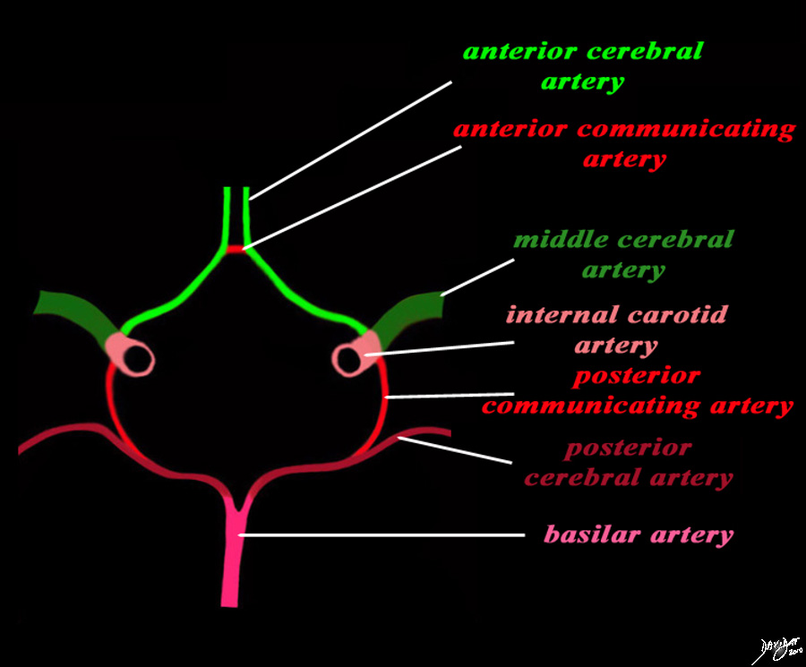
The diagram shows the main branches of the blood supply to the brain which includes the carotid and vertebro-basilar systems. These are the vessels that particpate in the formationofthe circle of Willis The carotid system supplies the brain from the internal carotid (salmon pink) – a branch of the common carotid which arises from the aorta. Its terminal bifuracation into the middle cerebral (dark green) and anterior green (bright green) are shown. The anterior communicating artery runs between the two anterior cerebrals (bright red) The basilar artery (pink) is formed by the two vertebral arteries and travel as a single artery over the upper medulla and the entire pons. Its terminal branch is the posterior cerebral artery (maroon). Each of the vessels contributes to the circle of Willis through communicating arteries. The vertebro-basilar system provides the posterior communicating arteries bilaterally and the carotid system provides the anterior communicating arteries via the anterior cerebral artery. Courtesy Ashley Davidoff MD Copyright 2010 All rights reserved 97194b13g04L01.91s |
The basilar artery runs in the basilar groove until it splits, giving both posterior cerebral arteries. During this journey, it provides the anterior inferior cerebellar artery and anterior superior cerebellar artery.

Vertebral Circulation – Lateral Projection |
| The lateral angiogram shows the vertebral artery feeding the basilar artery which in turn gives rise to cerebellar branches and and the posterior cerebral circulation which supplies posterior fossa structures and the occipital lobe. The detail of these vessels follows.
49416 Courtesy Ram Chavalli MD |

Capillary Phase – Posterior Circulation |
| The capillary phase of the vertebral injection provides insight into the perfusional segments that the posterior circulation feeds which includes structures of the posterior fossa, and structures of themid and hindbrain as well as the occipital lobe.
49417 Courtesy Ram Chavalli MD |

Vertebral Circulation – A-P Projection |
| The A-P projection of the right vertebral injection shows cross filling into the left vertebral, the basilar artery and the posterior cerebral artery. The posterior inferior cerebellar artery (PICA) is the first branch off the basilar artery but its origin off the basilar is slightly more difficult to appreciate. The origin and course of the the superior cerebellar artery is is easily seen and recognized as it arises just inferior to the larger posterior cerebral arteries which are the terminal branches of the basilar artery. In the A-P projection they assume a vertical course as they make their way to the occipital lobes.
49418 Courtesy Ram Chavalli |