White Matter
Copyright 2010
Definition
White matter shiny due to insulated sheaths
White Matter and Gray Matter
|
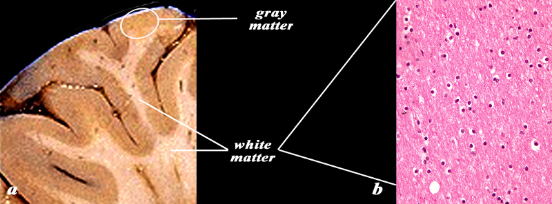
Histology 10X of the Gray Matter |
|
The image shows a gross specimen photographed to show the gyral pattern and the distinction between the rim of gray matter (light brown) and the white matter (cream colored) (a). The microsopic section (b), shows the histological equivalent of the gray matter as might be seen in the white circle consisting of a superficial layer of meninges and the deep layer of gray matter . A variety of cell types and sizes can be distinguished evn at this microscopic level. Image Courtesy of Thomas W.Smith, MD; Department of Pathology, University of Massachusetts Medical School. 97630bc.8 |
White Matter
|
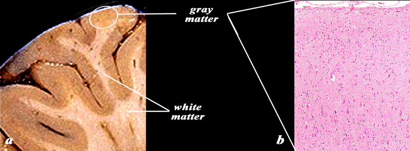
Histology of the White Matter (40X) |
|
The image shows a gross specimen (a) photographed to show the gyral pattern and the distinction between the rim of gray matter (light brown) and the white matter (cream colored). The microscopic section (b) is 40X magnified, and shows the histological equivalent of the white matter consisting mainly of oligodendrocytes characterized by cells with a white halo. No neurons are present Image Courtesy of Thomas W.Smith, MD; Department of Pathology, University of Massachusetts Medical School. 97630bc02.8 |
|
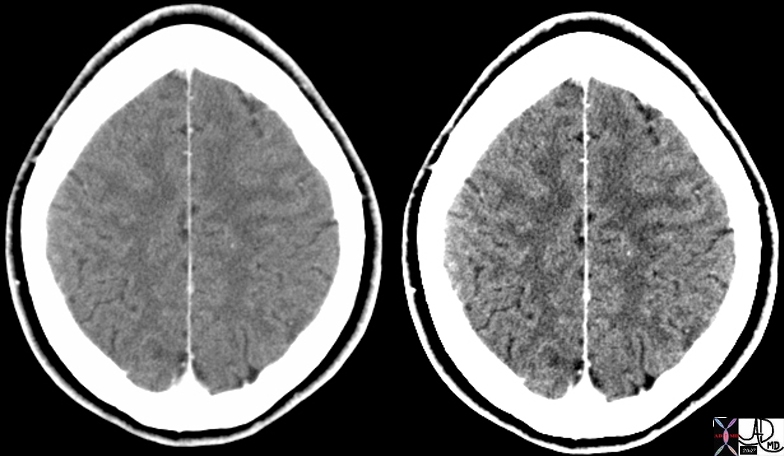
Normal Head CT Distinction Between Gray and White Matter |
|
The two images are form the same patient and the routine settings are shown on the left and the windows have been further narrowed on the right image in order to accentuate the difference between the gray and white matter. CT shows the white matter to be darker (blacker) than the gray matter which is a thin mantle of tissue on the edge of the brain. The ability to distinguish gray matter from white matter is a far easier task on MRI. Courtesy Ashley Davidoff MD copyright 2010 48757c01 |
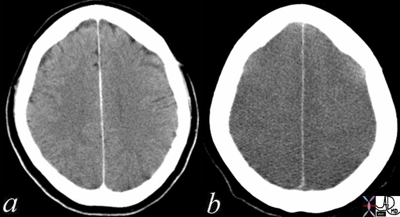
Normal – 4 months Prior and Folllowing Cardiopulmonary Arrest Loss of Gray White Differentiation |
|
The CT is from a 52 year old female who had a CT 4 months prior tto a cardiac arrest (a) showing subtle but definite difference in the attenuation difference between gray and white matter. After the cardiac arrest, she suffered anoxic injury to the brain resulting in extensive and global swelling of the brain and both gray and white matter have the same attenuation. The distinction between the two cannot be made and this finding is consistent with global anoxic injury. After 10-15 seconds of anoxia from cardiac arrest, loss of conciousness occurs and after 5 minutes of arrest, irreversible brain injury occurs. The gray matter of the brain, particularly the frontal lobes have highest metabolic needs. The occipital, parietal, and temporal lobes and basal ganglia and cerebellum have lesser needs. The brainstem has the lowest oxygen needs Courtesy Ashley Davidoff MD Copyright 2010 70134c01 |
|
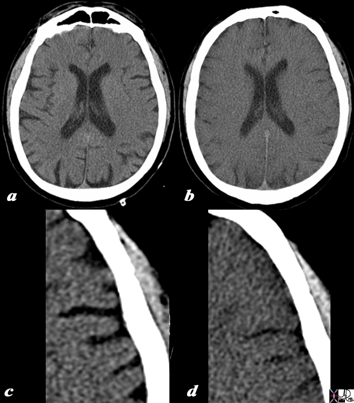
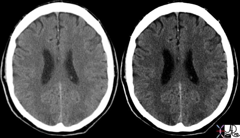
Normal (a,c) and 3 Days later after a Cardiac Arrest (b) |
|
The CT is from a 57 year old male who had a CT 3 days prior (a,c) to a cardiac arrest (b,d) showing subtle but definite difference in the attenuation difference between gray and white matter. After the cardiac arrest, he suffered anoxic injury to the brain resulting in extensive and global swelling of the brain and both gray and white matter have the same attenuation. The distinction between the two cannot be made and this finding is consistent with global anoxic injury and edema. The windows of (c) and (d) have been narrowed in order to accentuate the gray-white matter interface in c and show the graying out of the interface when the brain is edematous. c After 10-15 seconds of anoxia from cardiac arrest, loss of consciousness occurs and after 5 minutes of arrest, irreversible brain injury occurs. The gray matter of the brain, particularly the frontal lobes have highest metabolic needs. The occipital, parietal, and temporal lobes and basal ganglia and cerebellum have lesser needs. The brainstem has the lowest oxygen needs. Image Courtesy Ashley Davidoff MD Copyright 2010 All rights reserved 90637c02.8L01 |
|
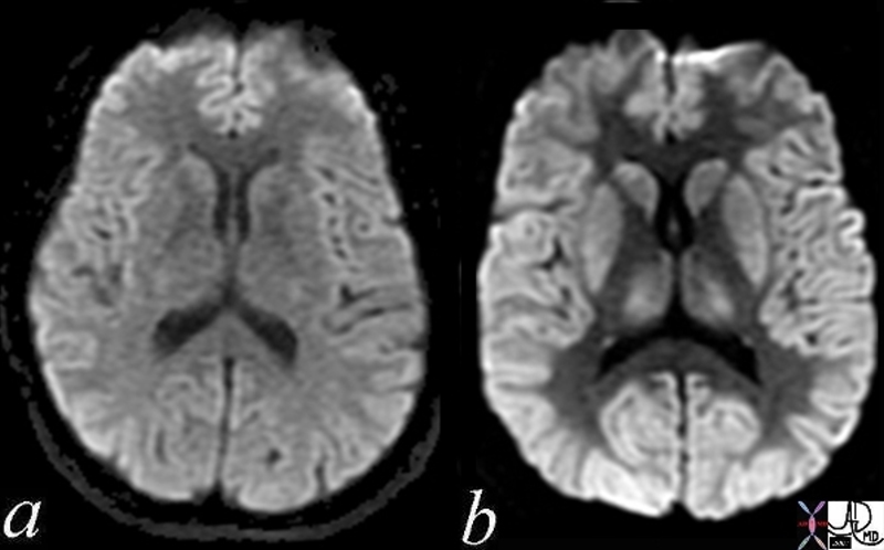
DWI – Normal (a) and Global Ischemia (b) |
|
The two images represent a diffusion weighted MRI sequence which measures Brownian motion of molecules. Image 9a) is normal. In acute infarction there is restricted Brownian motion of the affected area and the image can be manipulated to present this as a bright region. In this case the acute infarction or ischemia (b) is diffuse so the whole brain is relatively bright so that the white matter appears relatively dark (a) Courtesy Ashley Davidoff MD Copyright 2010 All rights reserved 49433c02.800 |
|
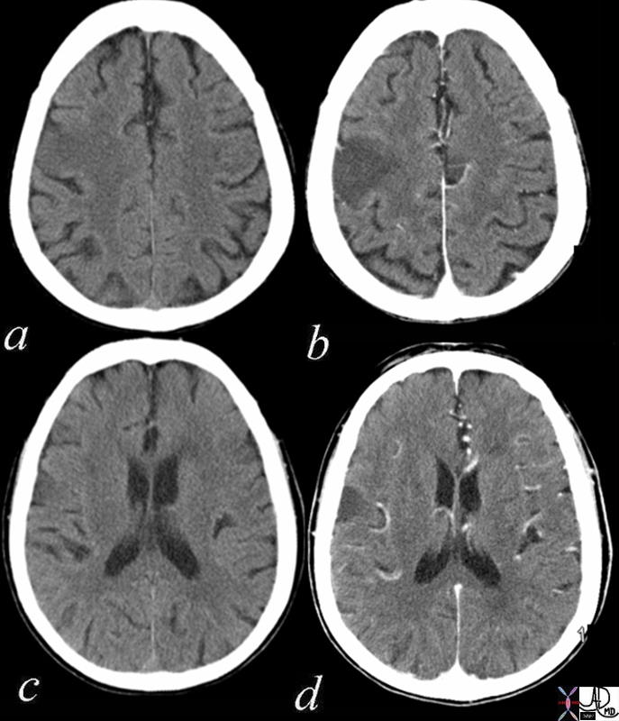
Acute Infarction Right Fronto-parietal Region |
|
The axial CT scan through the ventricles is from a 73year old female with acute left sided weakness and slurred speech. Images a nd c are without contrast and images b and d are following intravenous contrast. In the right frontoparietal region within the location of the pre and post central gyrus there is a focal hypodense region accentuated in the contrast study, with loss of the gray white differentiation, consistent with an acute infarction in the parietal lobe. Courtesy Davidoff MD Copyright 2010 71253c01 |
|
Subtle Occipital Lobe Loss of Gray White Differentiation |
|
This hypertensive patient presents with visual disturbance and the axial CT scan shows subtle changes in the occipital lobe characterised by vague low density regions. There is loss of gray-white matter differentiation consistent with acute ischemia. These findings are consistent with acute hypertensive encephalopathy Courtesyt Ashley Davidoff MD 73422 |
|
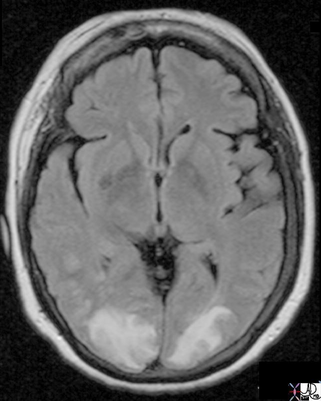
FLAIR Hyperintensity in the Occipital Lobes |
|
This hypertensive patient presents with visual disturbance and the MRI using a FLAIR sequence shows obvious FLAIR hyperintensity in the occipital lobes and extending into the parietal lobes. These findings are consistent with acute hypertensive encephalopathy and ischemia of the occipital lobes characteristic of acute hypertensive encephalopathy Courtesy Ashley Davidoff MD Copyright 2010 All rights reserved 73422 |
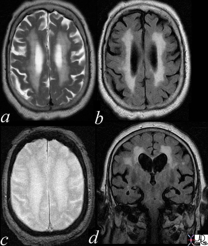
Microangiopathic Disease in the White Matter |
| The MRI is from an 82 year old femalewho shows increased water around in the white matter as seen inthe periventricular area on T2 weighted (a), FLAIR (b) , GRE (c), and FLAIR coronal images. The changes are reflective of age related disease of the smallest vessels at the distal end of the carotid and are said to reflct microangiopathic changes.
71472c04 Courtesy Ashley Davidoff MD |
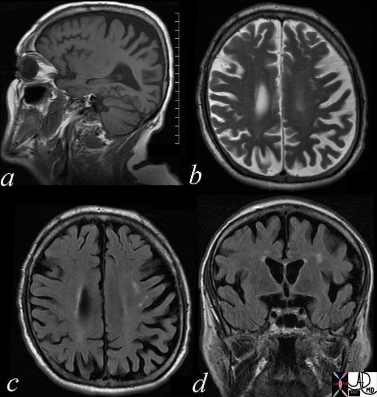
Lcune in the Corona Radiata on Left |
| 71609c01 brain cerebrum white matter corona radiata lacune chonic infarction a= T1 b= T2 weighted image c= FLAIR d= FLAIR coronal MRI Davidoff MD |

White matter fiber calcification |
| 48705c01 brain white matter fibers fibres fx calcified white matter fibers fx heavy calcification dystrophic calcification vs metastattic ddx Fabry’s disease hyperparathyroidism CTscan Courtesy Ashley Davidoff MD |
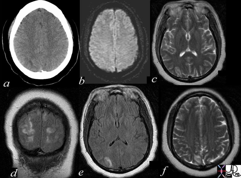
Posterior Reverrsible Encephalopathy |
| 72413c01.800 34 year old female with ecclampsia brain cortex and subcortical white matter parietal lobes occipital lobes frontal lobes distribution s consistent with posterior reversible encephalopathy syndrome a = CTscan b = DWI c T2 weighted image through lesions in occipital region d= FLAAIR coronal e = FLAIR axial f = T2 weightes with frontal lobe invlvement Davidoff MD |
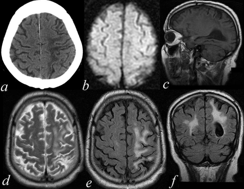
Chronic Ischemic Change in the left MCA territory – Fronto Parietal region |
| 71411c01.800 74 male with mental status changes brain cerebrum cerebral fx gliosis volume loss enlargement lateral ventricle on left encephalomalacia dx superior left frontal lobe parietal lobe in the territory of the left MCA left middle cerebral artery a= CTscan b = DWI – negative c= T1 weighted image d= T2 weighted image e = FLAIR f = FLAIR coronal MRI Davidoff MD |

Acute Cerebral Hemorrhage with Mass Effect and Rupture into the Ventricles |
| 72143c01 brain cerebral hemorrhage hemorrhagic CVA hematoma lateral ventricles 4th ventricle fourth ventricle mass effect gray matter white matter loss of the gray white interface cerebral edema blood CTscan CVA Davidoff MD |
|
10 year old with Leukodystrophy |
|
This MRI is from a 10 year old male with known leukodystrophy. The findings on the MRI of the brain (a,b) are characterised by significant changes in the white matter of both frontal lobes. (green arrow), characterised by T2 and FLAIR hyperintensity These findings are consistent with leukodystrophy. Courtesy James Donnelly MD Copyright 2010 All rights reserved 23431c01 |

|
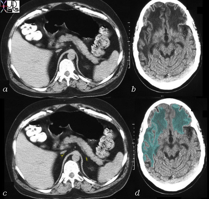
Adrenoleukodystrophy |
|
This CT is from a 26 year old male with known adrenoleukodystrophy. The findings on the CT of the brain (b,d) are characterised by significant changes in the white matter of both frontal lobes right parietal lobe and left parietal lobe to lesser extent. (green overlay) The adrenal glands (a,c) are barely visible but are overlaid in yellow in image (c). These findings are consistent with adrenoleukodystrophy. Courtesy Rebecca Schwartz MD Copyright 2010 All rights reserved 31464c04 |