
Thalamus
The Common Vein Copyright 2010
Definition
The thalamus is the gateway to the cerebral cortex. It is a paired organ and represents the main part of the diencephalon and subserves both motor and sensory function. It is structurally and functionally situated between the cortex and the midbrain.
It derives from the Greek word meaning chamber or bedroom
Structurally it is positioned in the middle of the brain. Each thalamus measures about 4 cms is almost ovoid in shape and lie on either side of the third ventricle. They have specific nuclei with diffuse projections to and from multiple regions of cerebral cortex. Histologically groups of myelinated fibers and neurons characterize the thalamus. It consists of 50-60 nuclii with well defined cortical connections
From a functional standpoint the thalamus acts as a translator for the cerebral cortex and the cortex acts to correct the input and suppress irrelevant information. It is the relay center for information processing with the forebrain. The thalamus plays a large role in the autonomic nervous system; regulating sleep and arousal. It may also play a role in vestibular function. It processes sensory and to lesser extent motor information and plays a large role in the autonomic nervous system regulating sleep and arousal. It may also play a role in vestibular function. Reciprocal connections to cortex are involved in conciousness.
Diseases include strokes, Korsakoff syndrome, and thalamocortical dysrhythmia.
Clinically thalamic dysfunction causes somatosensry dysfunction and or central pain on the opposite side of the body of the affected thalamus. It may on the other hand cause contralateral anaesthesia or hyperaestesia. From a motor point of view it may cause contralateral weakness or ataxia.mood swings and obsessive compulsive disorders.
Treatment of diseases is limited
|
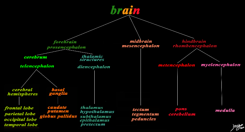
The Thalamus (teal green) Part of the Forebrain (Prosencephalon) Member of the Cerebrum (Telencephalon) Member of the Thalamic Family
|
|
The basic and simplest classification of the brain into forebrain midbrain and hindbrain is shown in this diagram and advanced to a more complex tree using the embryological and evolutionary terminologies. The forebrain consists of the cerebrum also called the prosencephalon, which contains the more advanced form of the brain and the thalamic structures which contain more basic structures. The cerebrum (telencephalon) itself consists of two cerebral hemispheres and paired basal ganglial structures. Each cerebral hemisphere will have gray and white matter distributed in the frontal parietal temporal and occipital lobe, with the basal ganglia being part of the gray matter deep in the cerebral hemispheres. The most important thalamic structures arising from the diencephalons include the thalamus itself and the hypothalamus. The midbrain (mesencepaholon) consists of the tectum tegmentum and cerebral peduncles. The hindbrain has two major branch points based on the evolutionary development. The pons and cerebellum(part of the metencephalon) are grouped and the medulla (part of the myelencephalon is the second branch. Courtesy Ashley Davidoff MD Copyright 2010 All rights reserved 97686.8s |
Thalamus – Forebrain Structure Epicentre of the Inverted C Rings |
|
The orange c shaped ring lies lateral and to some extent inferior to the lateral ventricles typified by the light blue ring and consists of the thalamus and basal ganglia including the globus pallidus, putamen, head of the caudate nucleus, tail of the caudate nucleus and the amydala. This diagram infers the concept of recurring inverted C shaped structures that are layered both from deep to superficial but also to some extent medial to lateral. Courtesy Ashley Davidoff MD Copyright 2010 All rights reserved 93907d13b05b02.8s |
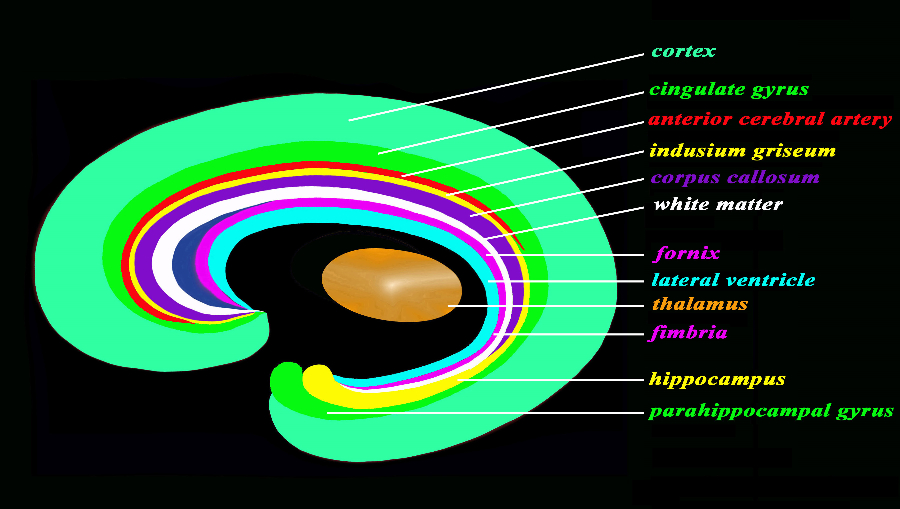
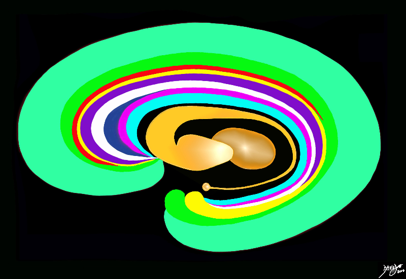
The Thalamus in the Middle of the Brain |
|
The forebrain has most of its components aligned in a series of inverted c- shaped rings starting from the outer cortex and advancing through a series of smaller inner rings with each intimately connected to the others. The thalamus appears diagramatically as the centre of these rings as seen from the sagittal view The outer and largest ring is the cerebral cortex, followed consecutively by the green ring which is the cingulate gyrus superiorly and the parahippocampal gyrus inferiorly. The red ring represents the distribution of the main portion of the anterior cerebral artery. Next is the yellow c which is the indusium griseum superiorly and the hippocampus inferiorly. This is followed by a the corpus callosum (purple) which enables the white ring of white matter to connect between hemispheres. The next ring is the thin pin kring whichrepresents the fornix superiorly and the fimbria inferiorly. The innermost ring (light blue) represents the lateral horns of the ventricular system The navy blue arrowheaded structure is the septum pellucidum Courtesy Ashley Davidoff copyright 2010 all rights reserved 93907d13a05b.9s |
|
The Orange Circle |
|
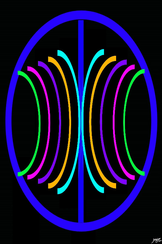
The anchoring concept of the brain in the axial plane is two cerebral hemispheres with a series of structures reminiscent of backward and downward facing “C’s” symettrically positioned around the center. The thalami and basal ganglia are part of the orange “circle” which lies along side the ventricles (light blue circle) Courtesy Ashley Davidoff MD copyright 2010 93914.3ka09.8sd01 |
|
Thalami and Caudate Nuclii |
|
The thalami (larger posterior orange structures) are diagramatically represented in close association with the tail of the caudate nucli posteriorly. They are positioned alongside the more centrally poisiiopned ventricles (light blue). Courtesy Ashley Davidoff MD copyright 2010 all rights reserved 93914.3ka04b01b02.8s |
|
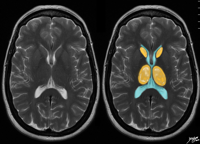
T2 Weighted MRI |
|
The next layer at this level through the large portion of the ventricles, (in this case the orange layer) the next bilateral backward “C’s are composed of the caudate nuclii anteriorly and the larger thalami posteriorly . Courtesy Ashley Davidoff MD copyright 2010 all rights reserved 94079cd05.8s |
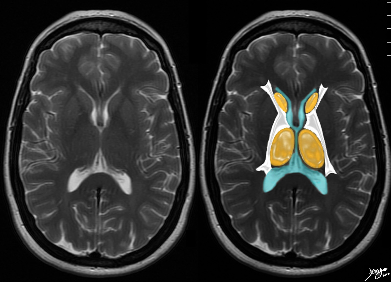
The Lateral Relations The Internal Capsule |
|
The axial T2 weighted MRI is taken through the level of the ventricles. The next outward layer is a white matter layer and at this level it represents the internal capsule brain Courtesy Ashley Davidoff MD copyright 2010 all rights reserved 94079cd06b01.81s |
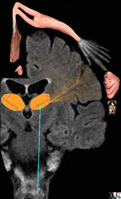
The Thalamus the Homunculus and the Somatosensory Cortex |
|
The diagram reflects the relative space each body part occupies in the somatosensory cortex by reflecting organs that have high density of sensory receptors and nerves as large organs and those with a lesser sensory apparatus as small organs. Hence the mouth lips, hands feet and genitalia have a large representation. The homunculus man (literally the “little man”) is the distorted figure drawn to reflect the concept of size of organ paralleling the size of the sensory innervation. Courtesy Ashley Davidoff MD Copyright 2010 38610b09.46k.8s |
Integrated Function with the Basal Ganglia and Cortex |
|
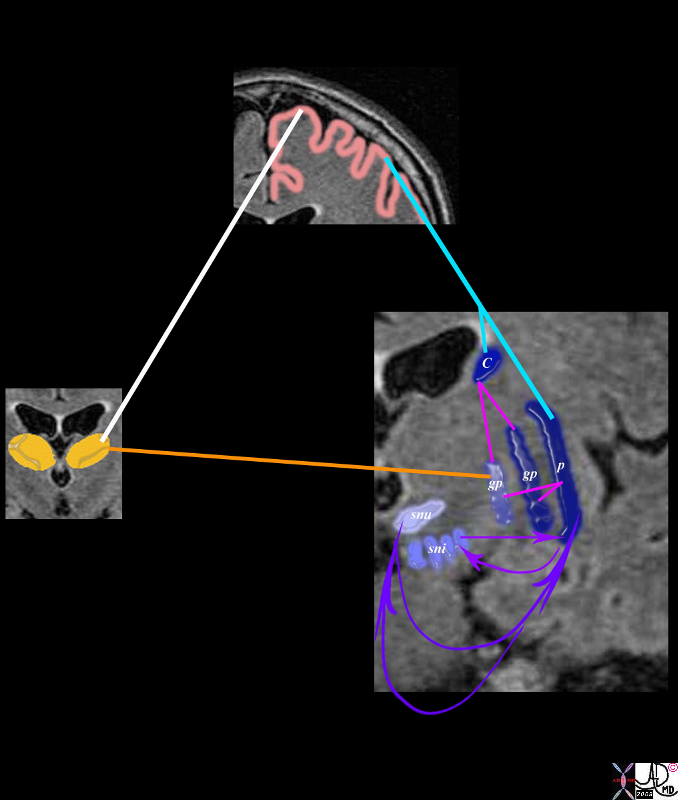
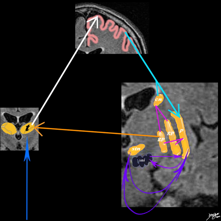
A simplified drawing adapted from a T1 weighted MRI showing the connections between the caudate nucleus (orange, the sensory cortex (salmon pink) and the basal ganglia is shown. After the stimulus has reached the sensory cortex for quantification and qualification it connects to the basal ganglia through the caudate nucleus and putamen. Each of these connect with the two parts of the globus pallidus (gp) which feed back to the thalamus. The caudate nucleus also feed back and forth to the substantia nigra (sni) and the subtalamic nucleus (snu) putamen= p caudate nucleus = cn globus pallidus = gp substantia nigra = sni subthalamic nucleus = snu Courtesy Ashley Davidoff MD Copyright 2008 38610d09e02.8s |
Thalamus as Part of the Reticular Activating System |
|
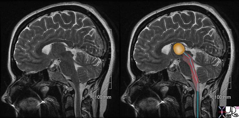
The reticular activating system (aka ascending reticular activating system) is a part of the brainconsidered to be the center of arousal and motivation. Structurally it lies betweent the medulla oblongata and midbrain and is connected to the thalamus. In turn al parts of the brain can be stimulated as a result including the cerbral cortex basal regions of the brain and the medulla Functionally it indirectly relates to our state of conciousness, and is involved with the control of the circadian rhythm, respiration, cardiac rhythms, and sexual function. In the instance of painthe RAS is activated by the C fibers and hence pain can arouse us from sleep through the RAS, can create a sense of urgency, and can cause changes in heart rate or respiration rate. Courtesy Ashley Davidoff MD copyright 2010 77059c01b01.8s |
The Thalamus as Part of the Limbic System |
|
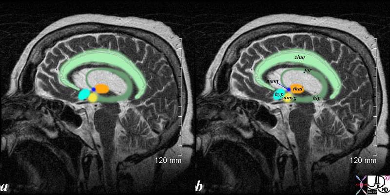
The sagittal T2 weighted MRI shows the limbic system which is a compound set of structures including the cingulate gyrus, (light green) cingulate cortex, hypothalamus (light blue) mamillary body (royal blue) fornix (darker green) hippocampus (darker green) amygdala (yellow) and thalamus (orange) Courtesy Ashley Davidoff MD copyright 2010 71430.85c01s |
Role of the Thalamus in the Pain Sensation
The thalamus is the gateway to the cerebral cortexand acts as a relay station to and from all parts of the spinal cord and brain and serves as an interpreter of information.
In the context of the pain process, it receives pain fibers (second order neurons) via the spinothalamic tract and directs them to the posterir medial and posterior lateral nuclii.
The thalamus translates pain signals of the 2nd order neurons and gives rise to the third order neurons that extend to the cortex. Awareness and localization of the pain is then achieved at the level of the cortex. The thalamus however is not merely a relay station for nociception but also plays a role in processing the stimulus.
The lateral pain system (venteroposteriorlateral VPL) projects via the lateral thalamic nuclei to somatosensory cortex where localization and duration of the stimulus is perceived. The medial pain system, (venteroposteriormedial VPM) projects through medial thalamic nuclei to the prefrontal and anterior cingulate cortices where the unpleasantness and emotional aspects of the pain is perceived.
Axons terminating in the lateral thalamus mediate discriminative aspects of pain (somatosensory cortex) including the originating body part. The fibers ending in the medial thalamus mediate the motivational and affective aspects relating for example to the emotional and memory of pain. These third order neurons travel to the prefrontal cortex, insular and cingulate gyrus which contribute to the emotion and memorization of pain experiences.
The Thalamus and networks inVolving the Paoin Sensation |
|
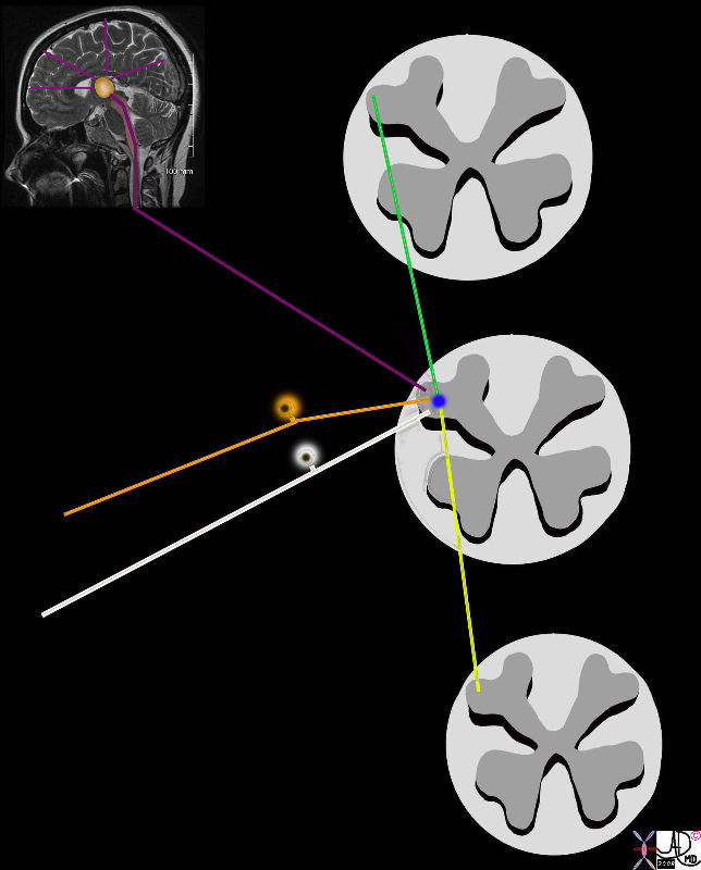
The orange fiber is one of many carrying the original pain stimulus as the first order neuron. It joins many nerves that synapse with the second order neuron in the dorsal horn, including the white A beta non nociceptor fiber, ascending (yellow) and descending (purple) tracts from other levels of the spinal cord, and descending tracts from the cortex, thalamus and other hgiher levels (green). Davidoff art copyright 2008 77533b01e05.8s |
Some of the Nuclii of the Thalamus Involved with Pain |
|
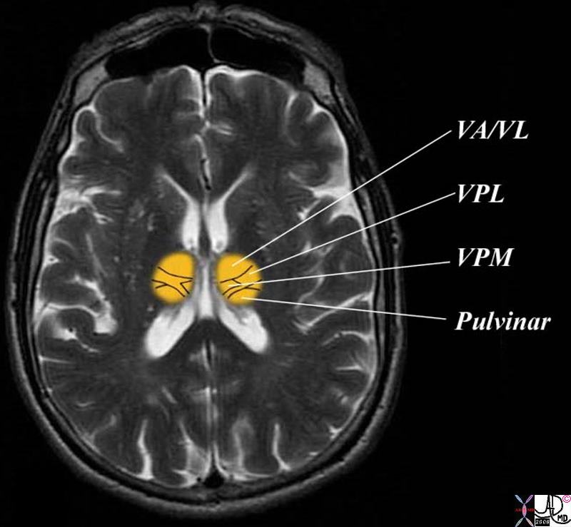
Thalamic pain centres involve the ventroposteriorlateral (VPL) and venteroposteromedial (VPM) nuclii, centeromedian and pulvinar nuclii. Courtesy Ashley Davidoff MD 38694c03b01.8s |
Acute Hemorrhage in the Thalamus |
|
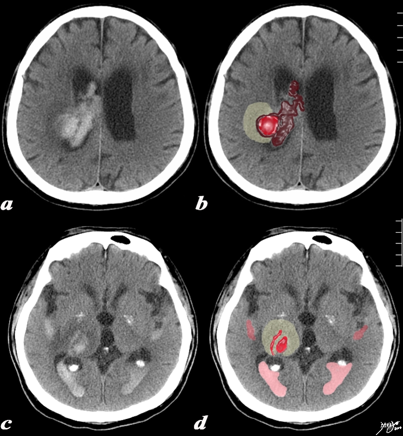
The CT is from a 77year old male with acute neurological deficit The epicenter of the disease is an acute hemorrhage in the right thalamus, bright red in (b,d) with extension of the clot into the ventricle (maroon). There is non clotted blood lying dependently in the occipital horns (dense on c) presenting as a CSF-blood level (light pink on black CSF) in (d). The hemorrhage is surrounded by a rim of edema (light yellow) as seen in b and d. Courtesy Ashley Davidoff MD Copyright 2010 All rights reserved 90461c03.8s |
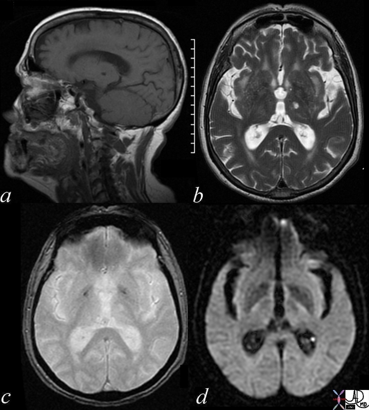
Hyperacute hemorrhage in the Right Thalamus |
|
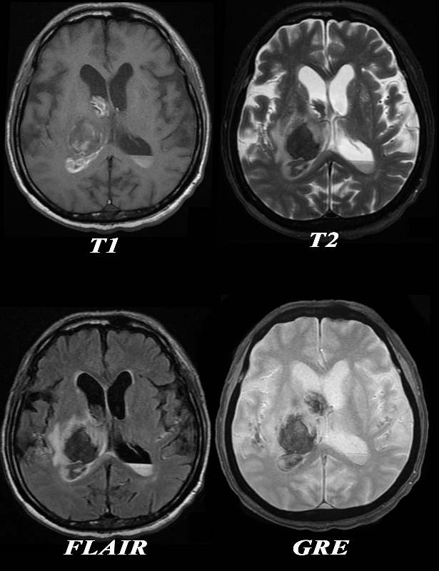
The MRI is from a 77year old male with acute neurological deficit The epicenter of the disease is a hemorrhage in the right thalamus. The T1 weighted image (upper left) shows mostly hypointense signal indicating the presence of deoxyhemoglobin. On the T2 weighted image the signal of the hematoma is also mostly hypointense confirming the hyperacute nature of the hematoma, surrounded by a ring of increased signal reflecting edema. There is a layering effect of blood in the posterior horn on the left The presence of deoxyhemoglobin dates the hemorrhage between minutes to hours The FLAIR image (c) shows the hyperacute hematoma as hypointense and the surrounding edema as hyperintense. The GRE image fails to reveal hemosiderin deposit in the periphery making chronic hemorrhage unlikely. The diagnosis of hyperacute intracerebral hemorrhage (minutes to hours) is therefore made by the presence of deoxyhemoglobin hypo/isointensity on the T1 weighted images and the hypointensity on T2 weighted. The suurounding edema is best seen on the T2 and FLAIR images. The extension into the ventricles is apparent on all sequences. Courtesy Ashley Davidoff MD Copyright 2010 All rights reserved 90484c04.8 |
Chronic Infarct in the Thalamus |
|
This series of 4 MRI images through the thalamus demonstrate a lacune in the left thalamus. Image (a) is a T1 weighted sagittal through the left thalamus and shows a hypointense defect, confirmed as a high intensity lesion on the T2 weighted image (b). The lesion is barely seen on the GRE image(c) and not seen on the DWI image(d). These findings are consistent with a chronic infrct in the left thalamus. brain cerebrum cerebral thalamus infarct chronic infarction circulatory MRI sagittal T1- focal hypointense T2 focal hyperintense GRE gradient echo – bright DWI diffusion weighted imaging Davidoff MD 70007c01 |
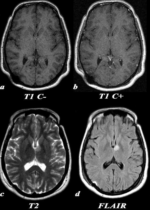
Low Grade Astrocytoma Left Thalamus |
|
A 31 year old female presented with transient episodes of numbness and tingling in her right face, arm and leg. MRI: T1 C- (a) No definite signal abnormality is identified in the left thalamic low grade glioma. T1 post (T1 C+ b) : Post contrast images demonstrate no enhancement in the region of the known mass. This is typical of a low grade glioma. T2 (c): This T2 weighted image shows a unilateral well circumscribed area of increased signal in the anterior left thalamus. There is no significant mass effect on the adjacent structures. FLAIR (d): This FLAIR image from her MRI demonstrates (with slightly better clarity than the T2 weighted image) a unilateral well circumscribed area of increased signal in the anterior left thalamus. There is no significant mass effect on the adjacent structures. . Courtesy Elisa Flower MD and Asim Mian MD 97664c.8 |
References
Netter Concise Neuroanatomy Lancet Student