
Copyright 2010
Introduction
The lateral surface of the temporal lobe is composed of the superior, middle, and inferior temporal gyri.
The superior temporal gyrus continues into the lateral sulcus.
Here it forms one of its walls.
Part of the superior temporal gyrus forms the temporal operculum.
The inferior temporal gyrus continues onto the inferior surface of the lobe.
The rest of the inferior surface is made up of the broad and often discontinuous occipitotemporal (fusiform) gyrus and the parahippocampal gyrus.
These are separated from one another by the collateral sulcus.
The occipitotemporal gyrus is partly in the occipital lobe and partly in the temporal lobe.
The parahippocampal gyrus is continuous with the cingulate gyrus around the splenium of the corpus callosum.
This is by the isthmus of the cingulate gyrus.
The anterior end of the parahippocampal gyrus turns backward and forms a medially directed bump called the uncus.
The superior border of the parahippocampal gyrus is the hippocampal sulcus.
Folded into the temporal lobe at the hippocampal sulcus is the hippocampus.
This is part of the limbic system.
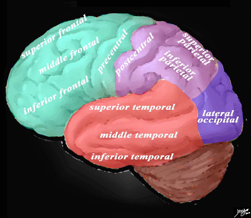
Overview of the Gyri of the Forebrain – Lateral External View |
|
The lateral view of the brain shows the frontal lobe (green) parietal lobe (light mauve), the occipital lobe (purple) and the temporal lobe (red) In this view the frontal lobe gyri that are visible are; superior frontal, middle frontal, inferior frontal and precentral gyri. The parietal gyri include the post central, superior parietal and inferior parietal. The occipital gyrus that is visible is the lateral occipital. The temporal lobe gyri include the superior, middle and inferior temporal gyri. Courtesy Ashley Davidoff MD Copyright 2010 83029d05g01.8s |