
The Common Vein Copyright 2010
Definition
The cerebral peduncles are tracts of fibres that run on the ventral surface of the midbrain. They are composed of white matter tracts that run between the brain and spinal cord including the the corticospinal tract fromn the internal capsule the corticobulbar tract, fibers from the substantia nigra.
The interpeduncular fossa lies between the cerebral peduncles and the interpeduncular cisterns lie anteriorly in the subarachnoid space.
Axial Plane
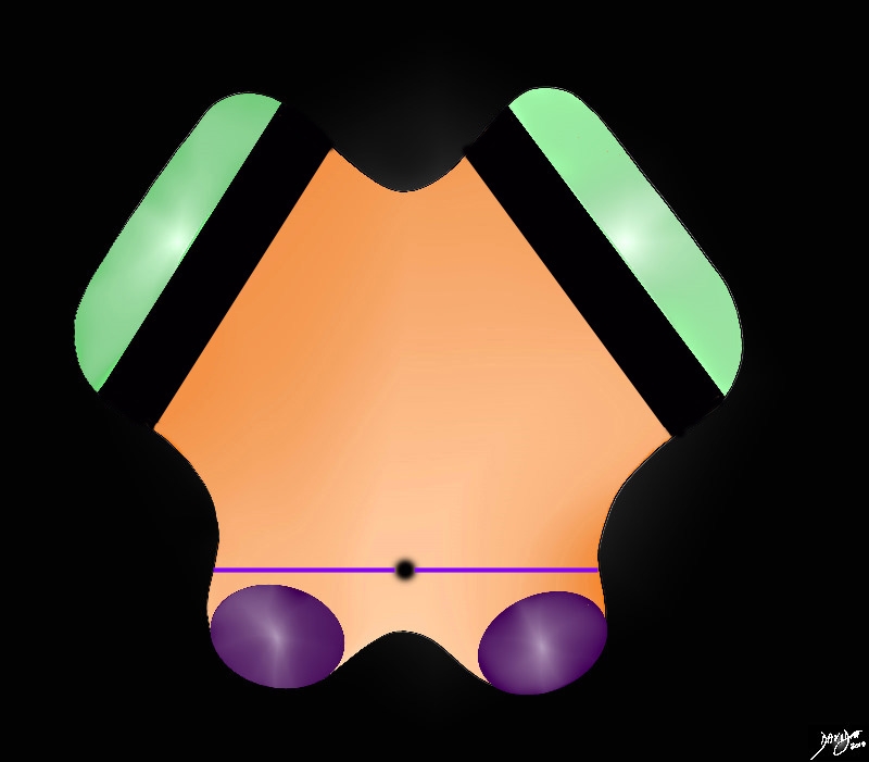
Cerebral Peduncles (green) |
|
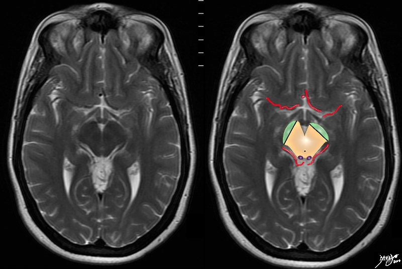
The anterior border of the midbrain incorporates the cerbral peduncles(green), and the substantia nigra (black – just posterior to the peduncles. Between the substantia nigra and the aqueduct is an area of the midbrain called the tegmentum (floor of the midbrain) The posterior end of the midbrain is bordered by the colliculi (purple) in the tectum (roof) of the midbrain. Courtesy Ashley Davidoff MD copyright 2010 all rights reserved 94074b09b05b.83s |
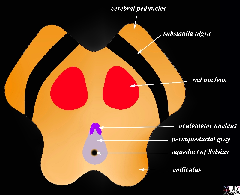
Cerebral Peduncles in Axial Projection |
|
The midbrain in transverse plane illustrates the component structures, with the image reminiscent of the face of a baby pig.. Anteriorly the cerebral peduncles are followed by the substantia nigra, and the collilculi The anterior border of the midbrain incorporates the cerebral peduncles, and the substantia nigra (black – just posterior to the peduncles). Between the substantia nigra and the aqueduct is an area of the midbrain called the tegmentum (floor of the midbrain) Within the tegmentum other structures include red nuclii, oculomotor nuclii, periaquaductal gray, and the aqueduct of Sylvius which is border forming between the tegmentum anteriorly and the tectum (roof) posteriorly. The posterior end of the midbrain is bordered by the colliculi in the tectum. Courtesy Ashley Davidoff MD copyright 2010 all rights reserved 94074b08a06bL.9s |
|
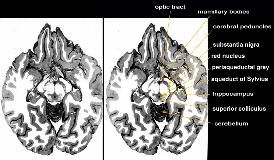
Axial Anatomic Specimen through the Midbrain Periaqueductal Gray Around the Aqueduct of Sylvius |
|
The anatomic section is an axial slice through the brain showing the structures of the midbrain. The most anterior structures of the midbrain are the cerebral peduncles followed by the substantia nigra, red nucleus, periaquaductal gray (PAG or central gray), aqueduct of Sylvius and finally the superior colliculus. Structures that are related to the midbrain anteriorly are the mamillary bodies and the optic tract. The hippocampus is positioned posterolaterally and the superior aspect of the cerebellum is seen posteriorly. Courtesy Department of Anatomy and Neurobiology at Boston University School of Medicine Dr. Jennifer Luebke , and Dr. Douglas Rosene 97140b03c.9 |
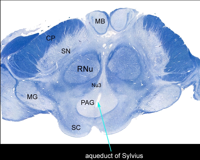
Whole Mount of the Midbrain in Axial Projection |
|
The mounted and stained midbrain in transverse plane illustrates the component structures. The anterior border of the midbrain incorporates the cerebral peduncles (CP), and the substantia nigra (SN). Between the substantia nigra and the aqueduct (teal arrow) is an area of the midbrain called the tegmentum (floor of the midbrain) Within the tegmentum other structures include red nuclii (RNu), oculomotor nucleus (Nu3), periaquaductal gray (PAG), and the aqueduct of Sylvius which is border forming between the tegmentum anteriorly and the tectum (roof) posteriorly. The posterior end of the midbrain is bordered by the colliculi in the tectum. Note also the paired mamillary bodies anteriorly (MB) and the medial geniculate body (MG) posterolaterally Courtesy Department of Anatomy and Neurobiology at Boston University School of Medicine Dr. Jennifer Luebke , and Dr. Douglas Rosene 98483L.8 |
|
The Cerebral arteries Characteristically Surround the Midbrain |
|
The MRI T2 weighted series now focuses on another very characteristic feature of the midbrain in that it is surrounded by the anterior and posterior cerebral circulations, and in this case portions of the arteries are seen with parts of the middle cerebral seen anteriorly and parts of the posterior cerebral seen posteriorly Davidoff art Courteys Ashley Davidoff MD copyright 2010 all rights reserved 94081.4kc02.8s |
Sagittal Plane
|
Basic Divisions of the Midbrain Tegmentum and Tectum Cerebral Peduncles, Cerebral Crura and Colliculli
|
|
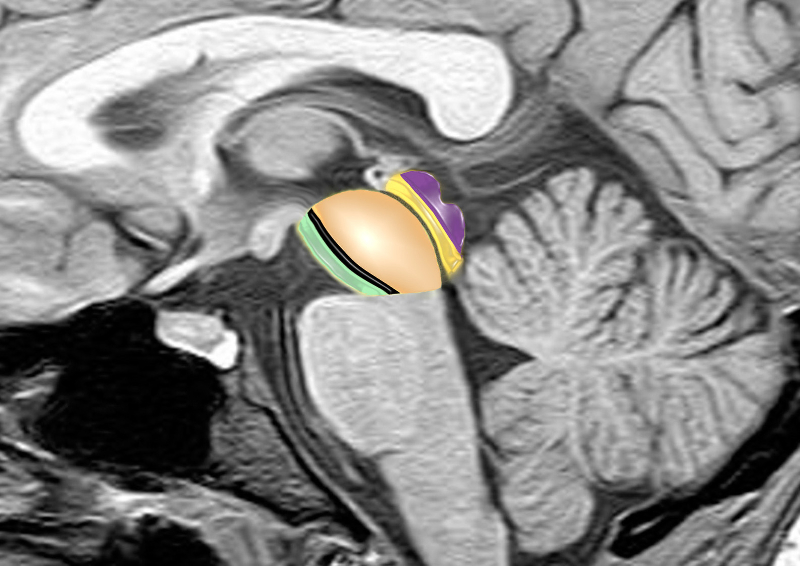
The midbrain consists of a larger anterior portion consisting of the cerebral peduncles, (green) the substantia nigra (black) and the tegmentum (light orange) that extends to the aqueduct (thin gray line) The posterior portion called tectum (orange) contains the colliculi (purple) which are the most posterior portions of the midbrain. brain anatomy neuroanatomy midbrain tegmentum colliculi tectum cerebral peduncle aqueduct of Sylvius conceptual diagram MRI principles Image provided by Philips Medical Systems Enhanced by Davidoff art Courtesy Ashley Davidoff MD 92141.3kb01ba02b02.8s |
Interpeduncular Cistern |
|
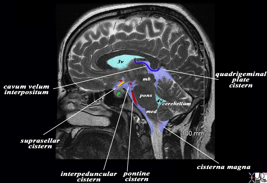
The overlay outlines the basal cisterns in light blue, and the ventricles (teal blue), and a variety of cerebral structures including the midbrain (mb) pons, medulla (med) and cerebellum. A variety of blood vessels (overlaid in blue and red are related to the basal cisterns. The image is T2 weighted sagittal MRI sequence from a patient with a macroadenoma of the pituitary (green p). Starting inferior and anterior we follow the pontine or prepontine cistern associated with the basilar artery, the interpeduncular cistern which is an anterior to the peduncles of the midbrain, the suprasellar cistern (above the pituitary – green p), and the cavum interpositum. Posteriorly the quadrigeminal plate cistern lies behind the colliculi and finally the cisterna magna which surrounds the inferior aspect of the medulla and cerebellum and merges with the subarachnoid space around the cord. The peripheral dural sinuses include the superior sagittal sinus, the laterally placed junction of the transverse sinus and straight sinus, and the confluens of the inferior sagittal sinus which runs on the inferior surface of the falx and the straight sinus which lies on the posterior and superior surface of the tentorium overlying the cerebellum. Image Courtesy Ashley Davidoff MD Copyright 2010 All rights reserved 98287b03L02b.9s |
Interpeduncular Cistern on CT |
|
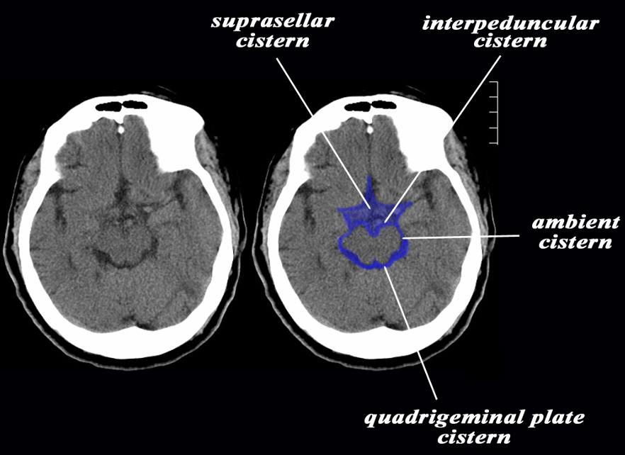
The axial CTscan through the midbrain shows the suprasellar cistern with a stellate shape anteriorly and enclosing vessels of the circle of Willis. Posteriorly the suprasellar cistern fuses with the interpeduncular cistern anteriorly, the ambient cisterns laterally, and quadrigeminal plate cisterns posteriorly. Image Courtesy Ashley Davidoff MD Copyright 2010 All rights reserved 94013b.9s |
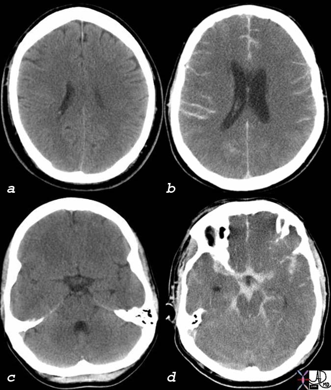
Subarachnoid hemorrhage Affecting Interpeduncular Cistern (d) |
|
The CT scan of this 52 year male is without contrats and reveals findings consistent with a subarachnoid hemorrhage likely caused by rupture of an aneurysm. In image b high density blood is seen in the sulci and in the interhemispheric fissureand in image d hemorrhage is seen in the stellate shaped suprasellar cistern and in the interpeduncular cistern of the midbrain, ambient cisterns, and likely in the aqueduct. Courtesy Ashley Davidoff MD 76059c01 76059c02 76059c03 76059c01 |