
Forebrain
Sagittal or Lateral View
The Common Vein Copyright 2010
The Forebrain
The forebrain is the largest and most complex of the 3 major parts of the brain, the two others being the midbrain and hindbrain.
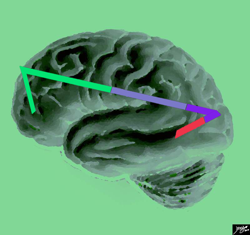
The Basic Vectors of the Forebrain |
|
The stick diagram defines the basic vectors of the forebrain, with anterior being to the left and posterior being to the right. This artistic rendition of the brain reflects the vectors of the major parts of the brain with the stick diagram overlaid over a sagittal external view of the brain. In the stick diagram, the forebrain has now been divided into the frontal lobe (bright green), parietal lobe (light mauve) occipital lobe (purple) and temporal lobe (red). The basal ganglia and the thalamic structures are not appreciated on this external view. The basal ganglia and the thalamic structures which complete the forebrain are not appreciated on this external view. Courtesy Ashley Davidoff copyright 2010 all rights reserved 83029e04.83s Davidoff art Courtesy Ashley DAvidoff MD copyright 2010 93887b03b01.8s |
The sulci and gyri can be quite confusing and so for simplicity we will start by defining the border forming sulci including the;
central sulcus that defines the border between the frontal lobe and parietal lobe
Sylvian fissure (aka lateral fissure) that separates the temporal lobe from the frontal and parietal lobe
parieto-occipital fissure that separates the parietal lobe from the occipital lobe
The border between the occipital lobe and the tempral lobe is ill defined
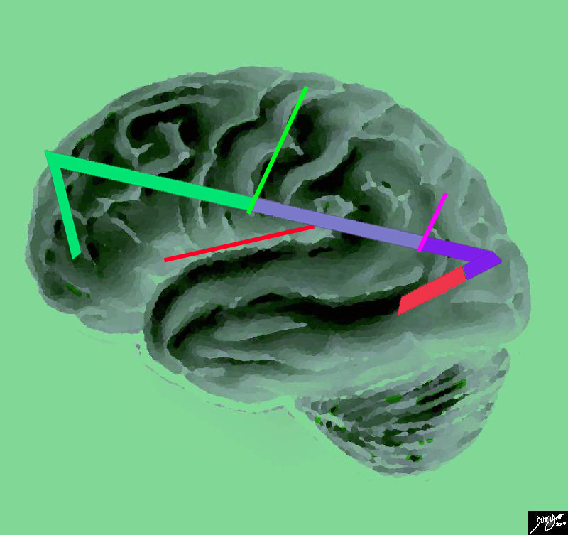
Major Border Forming Sulci and Fissures of the Forebrain |
|
This artistic rendition of the brain reflects the vectors of the major parts of the brain revealing the major border forming fissures and sulci. The central sulcus (bright green line) divides the frontal lobe from the parietal lobe (light mauve). The region of the parieto-occipital fissure (pink line) divides the parietal lobe from the occipital lobe (purple) and the Sylvian fissure (thin red line)divides the temporal lobe from the frontal and parietal lobe. Courtesy Ashley Davidoff copyright 2010 all rights reserved 83029e04.87s |
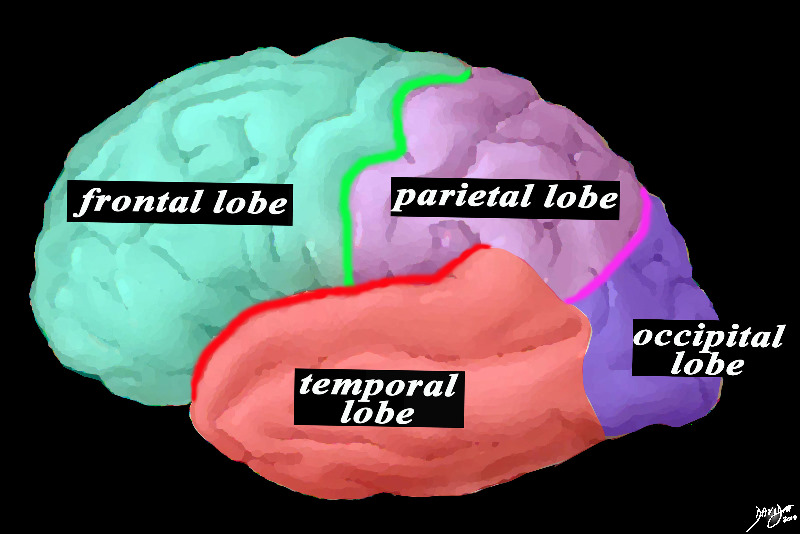
The parts of the Forebrain and the Borders |
|
This is a diagram looking at the forebrain from the side showing the anteriorly placed frontal lobe, separated from the inferiorly placed temporal lobe by the Sylvian fissure (red), and separated from the parietal lobe by the central sulcus (green). The parietal lobe is also separated from the temporal lobe by the Sylvian fissure and from the occipital lobe by the parieto-occipital fissure (pink). The basal ganglia and the thalamic structures which complete the forebrain are not appreciated on this external view. Courtesy Ashley DAvidoff MD copyright 2010 83029d13b01.8s |
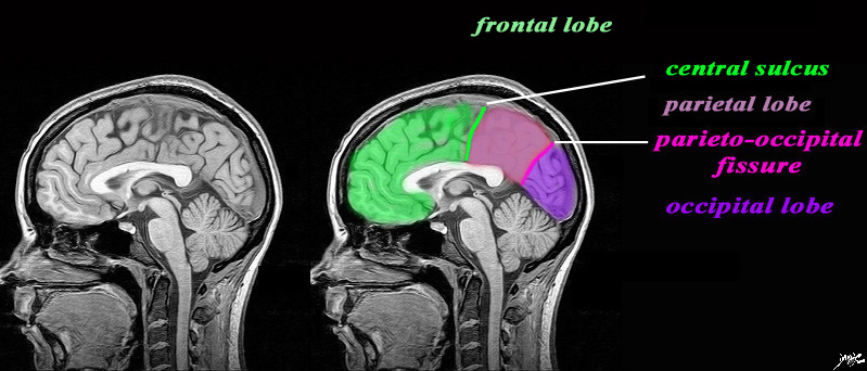
Deeper in the Brain – Sagittal View |
|
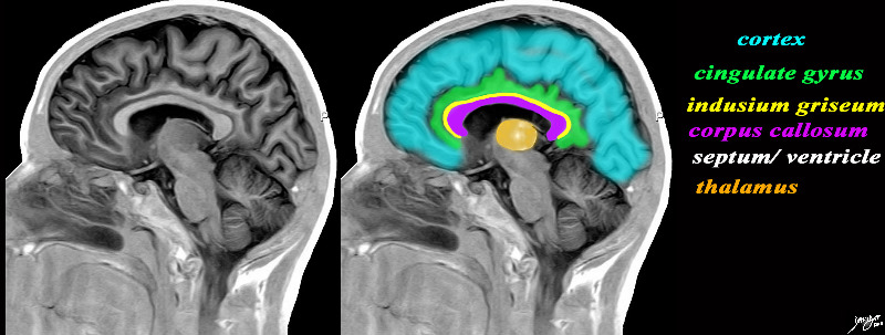
The sagittal image is deeper in the brain near the midline through the interhemispheric fissure, and is intended to demonstrate the the parieto-occipital fissure (pink) in order to define the border between the parietall and occipital lobe. The central sulcus (pink) has also been inferred in to demonstrate the border between the frontal lobe and parietal lobe. It is not usually seen in this projection. The Sylvian is also not usually seen in this projection. What we do start to appreciate are the layers of inverted c shaped structures. MRI 3D FFE sag mid Br Courtesy Philips medicaL systems rendered by Davidoff art copyright 2010 92141c01label.82s |
As we delve beyond the surface and into a midline sagital view we appreciate that the vector or form of the forebrain from superior to inferior is more like a series of inverted parallel – c shaped structures.
Forebrain A series of C rings |
|
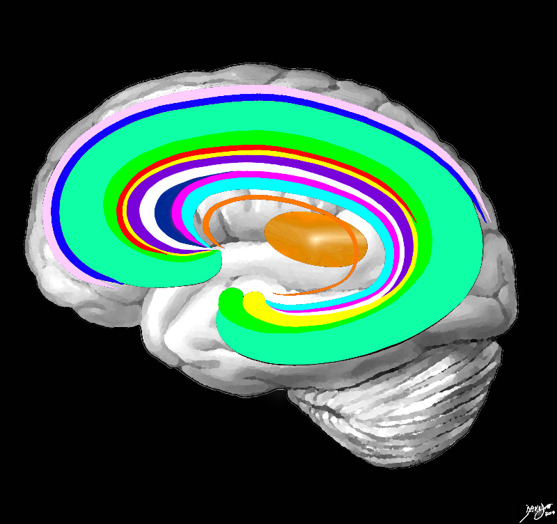
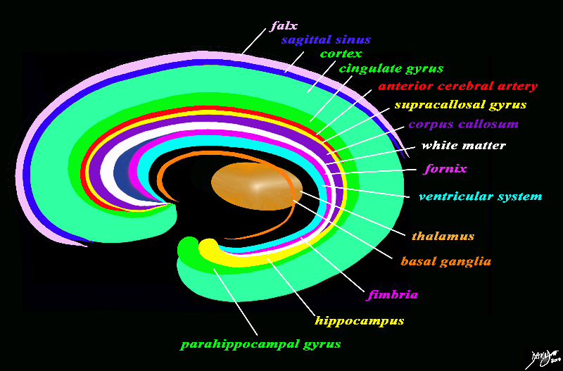
The forebrain has most of its components aligned in a series of inverted c- shaped rings starting from the outer membranes that culminate in the falx (pink), then extending inward smaller inner rings with each intimately connected to the others. The thalamus (dull orange) appears diagrammatically as the centre of these rings as seen from the sagittal view The outer ring is the falx (pink) followed by the sagittal sinus (blue) cerebral cortex (light green), cingulate gyrus (bright green) superiorly which becomes the parahippocampal gyrus inferiorly. The red ring represents the distribution of the main portion of the anterior cerebral artery. Next is the yellow ring which is the supracollosal gyrus (indusium griseum) superiorly and the hippocampus inferiorly. This is followed by the corpus callosum (purple) which enables the white ring of white matter to connect between hemispheres. The next ring is the thin bright pink ring which represents the fornix superiorly and the fimbria inferiorly. The innermost ring (light blue) represents the lateral horns of the ventricular system. The basal ganglia run just lateral to the lateral ventricles. The navy blue arrow headed structure is the septum pellucidum. Courtesy Ashley Davidoff copyright 2010 all rights reserved 83027b04g03.8s |
Some of the Inverted C-Shaped Structures |
|
The sagital view of the brain reflects some of the inverted c-shaped rings including the cortex, cingulate gyrus, indusium griseum, corpus callosum, septum pellucidum/lateral ventricles, and the thalamus which is central. Davidoff art Image Courtesy of Philips Medical Systems 92170c01b01b01.8s
|
Introduction to More Complex Neuroanatomy terms |
|
The forebrain has most of its components aligned in a series of inverted c- shaped rings starting from the outer membranes that culminate in the falx (pink), then extending inward smaller inner rings with each intimately connected to the others. The thalamus (dull orange) appears diagrammatically as the centre of these rings as seen from the sagittal view The outer ring is the falx (pink) followed by the sagittal sinus (blue) cerebral cortex (light green), cingulate gyrus (bright green) superiorly which becomes the parahippocampal gyrus inferiorly. The red ring represents the distribution of the main portion of the anterior cerebral artery. Next is the yellow ring which is the supracollosal gyrus (indusium griseum) superiorly and the hippocampus inferiorly. This is followed by the corpus callosum (purple) which enables the white ring of white matter to connect between hemispheres. The next ring is the thin bright pink ring which represents the fornix superiorly and the fimbria inferiorly. The innermost ring (light blue) represents the lateral horns of the ventricular system. The basal ganglia run just lateral to the lateral ventricles. The navy blue arrow headed structure is the septum pellucidum. Courtesy Ashley Davidoff copyright 2010 all rights reserved 93907d13ag03.8s |
The hemispheres have a form of a triangular prism , with 2 extremities, 3 faces and 3 borders. The anterior extremity is called the frontal, the posterior is called occipital.
The internal face, plane and vertical, limiting in each side, the interhemispheric fissure. The external face, convex, is in relation with the bony cranium. The inferior face is irregular. It has, in the junction of the anterior ¼ with the remainder ¾, a profound curved fissure, that is the initial portion of the Sylvian fissure. Ahead of this fissure, in the internal 1/3, a tract – the olfactory tract – is seen, that ends ahead in the olfactory bulb. Behind, the tract is originated by an internal white root and by an external white root. The former heads internally; the latter towards the hippocampal circumvolution. Behind the Sylvian fissure, the inferior face looks similar to a kidney whose hilum is directed inwards. The anterior portion forms the temporal pole of the cerebrum.
In terms of interhemispheric structures, above, there is a white formation called corpus callosum. Below, near the base, the optic chiasm if found; lateral to it we will find the anterior perforated substance. Behind the optic chiasm, the tuber cinereum, pituitary stalk, pituitary body, mamillary bodies and posterior perforated substance are found.
Corpus callosum, with a quadrilateral shape, measures between 8 to 10 cm in the longitudinal dimension; it has about 18 mm of transverse diameter in the superior face / 30-40 mm in the inferior face and 10-15 mm of height. Its superior face relates to the cerebral falx, cingulated gyrus and the pericallosal arteries. White and gray tracts can be seen. The inferior face relates with the fornix and septum pellucidum in the midline, and with the lateral ventricles in both sides. The anterior extremity has a curved shape, forming the genum, its most anterior part, the “peak”, being called the rostrum. The posterior segment is free and rounded, being termed splenium. The posterior angles form fiber radiations called the forceps major of the corpus callosum which bend sharply backward into the occipital lobe of the cerebrum. Similarly, another set of fibers which are more external are called the tapetum. The anterior angles also form fiber radiations which bend forward toward the frontal pole of the cerebrum; this is called the forceps minor.
The optic chiasm is made of white substance, having a quadrilateral shape, located behind the genum of the corpus callosum. Its inferior face rests over the hypophysis. Its superior face is connected to the base of the cerebrum thorough a very thin lamina consisting of gray matter, the lamina terminalis. The two anterior angles originate the optic nerves; the two posterior angles give rise to the optic tracts. These head posteriorly, surround the crus cerebri, later dividing in 2, which terminate in the internal and external geniculate bodies of the thalamus.
The tuber cinereum is a sheet of gray substance that occupies the space between the optic chiasm, the optic tracts and mamillary bodies. Its superior face is part of the third ventricle, being thus covered by ependymum.
The pituitary stalk is a small column of gray substance that begins in the most prominent part of the tuber cinereum down to the pituitary body. Its center in indented, which is due to a prolongation of the third ventricle.
The pituitary body, also called hypophysis, is a small elliptic mass suspended in the inferior extreme of the pituitary stalk.
The are 2 mamillary bodies. They are 2 white prominences, located in the internal part of the crux cerebri, behind the tuber cinereum.
The fornix is a lamina of white matter located immediately below the corpus callosum. It has a shape of a triangle with its base being posterior, being fused to the corpus callosum in the posterior third. Ahead of this part, it is connected to the same structure through the septum pellucidum, which is a vertical lamina situated in the midline. On each side of the septum pellucidum, the superior face of the fornix constitutes the floor of the lateral ventricles.