
The Common Vein Copyright 2010
Definition
Astrocytes are the largest cells of the glial tissue. They are named after their star shape.
Astrocytic processes form the glial membranes. These line the cerebral ventricles and central canal of the spinal cord. Astrocytes also store glycogen in their cytoplasm, which can be broken down to glucose and released into the surrounding neurons in response to the action of noradrenaline.
Astrocytes participate, along with microglia, in phagocytic activity, removing traces of nerve tissue, from degenerating axon synaptic terminals. After the death of neurons by pathological processes, astrocytes proliferate and fill the space created, a phenomenon known as replacement gliosis. They are very sensitive to changes in extracellular potassium and serve as buffers to prevent depolarization of neighboring neurons when the extracellular concentration of this ion increases.
The astrocytic processes differ from neurofilaments in that they are denser, have smaller diameter and different protein composition. Its key component is the protein glial fibrillary acidic, traditionally associated with nutritive support to neurons.
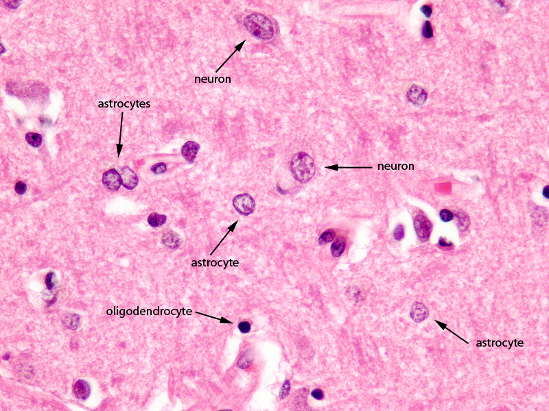
The Cerebral Cortex 60X Magnification |
|
The image shows a histological section of the brain at 60X magnification. The section shows the 3 of the common basic types of brain cells; The largest is the neuronal cell which is only found in gray matter. They are 10 times larger but less common than the second type of cell called the glial cell. The astrocyte is the second largest cell and often lies in close association with the blood vessels. In this section the astrocyte is exemplified. The third type of cell seen in this section, also a glial cell is the oligodendrocyte. It is smaller than the other type of glial cell the astrocyte , has a darker nucleus and it is responsible to myelinate the white matter. It is the predominant cell of white matter. Courtesy of Thomas W.Smith, MD; Department of Pathology, University of Massachusetts Medical School. 97801 |
They are classified according to their processes.
-Fibrous Astrocytes: with fine processes, found mainly in the white matter interposed between the fascicles of nerve fibers. They surround blood vessels through their processes, thus contributing to the blood-brain barrier.
-Protoplasmic Astrocytes: with few short and thick extensions, also surround blood vessel walls. Found in the pia mater.and mainly in the gray matter between the neuron bodies.
The human brain contains roughly 84.6 billion glia and 86.1 billion neurons. (Azevedo)
Most cerebral cortex glia are oligodendrocytes (75.6%) then astrocytes (17.3%) and least for microglia (6.5%) (Pelvig)
The amount of glial celss that are found in brain tissue is related to brain size
| animal | % glial cells |
| nematode | few glia |
| fruitfly | 25% |
| mouse | 65% |
| human | 90% |
| elephant | 97% |
(Allen)
References
Allen NJ, Barres BA. (2009). Neuroscience: Glia – more than just brain glue. Nature. 457(7230):675-7. PMID 19194443
Azevedo FA, Carvalho LR, Grinberg LT, Farfel JM, Ferretti RE, Leite RE, Jacob Filho W, Lent R, Herculano-Houzel S. (2009). Equal numbers of neuronal and nonneuronal cells make the human brain an isometrically scaled-up primate brain. J Comp Neurol. 513(5):532-41. PubMed
Pelvig DP, Pakkenberg H, Stark AK, Pakkenberg B. (2008). Neocortical glial cell numbers in human brains. Neurobiol Aging. 29(11):1754-62.(figures given are those for females) PubMed