The Common Vein Copyright 2010
Definition
The frontal gyri are the ridges of brain tissue seen on the surface of the frontal lobe that are separated by fissures and sulci consisting of mounds of gray and white matter that have been thrown into folds enabling an increase in the surface area of the brain in order to optimise space and hence optimise function.
The gyri of the frontal lobe structurally have characteristic morphological features that allow them to be recognized, named and attributed certain functions.
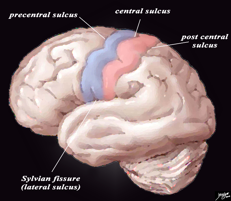
The precentral gyrus is part of the frontal lobe and lies in front of the central sulcus and is responsible for motor function for example; It runs at an almost vertical orientation, and runs parallel to the central sulcus and the postcentral gyrus.
|
The Precentral Gyrus (blue) and Postcentral Gyrus (pink) |
|
The lateral view of the external brain shows the most easily recognized gyri – the precentral gyrus motor cortex (blue) and the post central sensory gyrus (pink) surrounded by the sulci. There are 4 important central sulci (aka fissures), 3 of which are vertically oriented and 1 of which is horizontal. The vertical sulci include the central sulcus, precentral sulcus and post central sulcus. The central sulcus is important because it separates the frontal lobe from the parietal lobe. The post central sulcus is important because together with the central sulcus it forms the borders of the post central gyrus (pink) which is the sensory cortex. The precentral sulcus together with the central sulcus forms the borders post central gyrus (blue) which is the motor cortex. The Sylvian fissure or lateral sulcus runs horizontally and separates the temporal lobe below from the frontal lobe and parietal lobe above. Courtesy Ashley Davidoff copyright 2010 all rights reserved 83029b01b01b02g01L.8s |
The frontal gyri are best appreciated as they are seen from the lateral examination of the brain and from a sagittal view of the brain
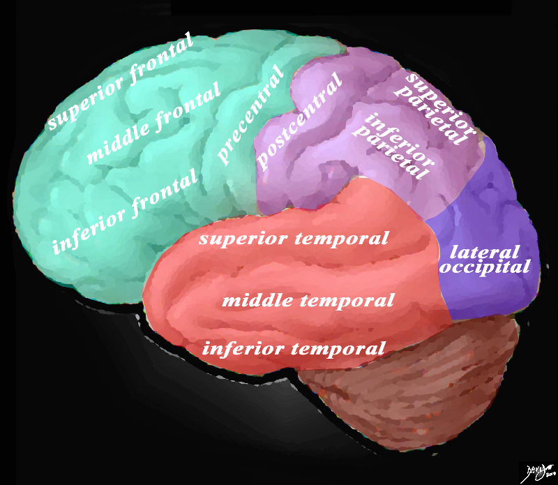
From the lateral aspect the most anterior gyri organized from superior to inferior and lying almost in the horizontal plane and parallel to each other are the superior, middle and inferior frontal gyri.
The superior frontal gyrus continues around the front of the brain toward the medial surface to reach the cingulate sulcus.
The inferior frontal gyrus is divided into 3 parts; opercular, triangular and orbital.

Overview of the Gyri from the Lateral External View |
|
The lateral view of the brain shows the frontal lobe (green) parietal lobe (light mauve), the occipital lobe (purple) and the temporal lobe (red) In this view the frontal lobe gyri that are visible are; superior frontal, middle frontal, inferior frontal and precentral gyri. The parietal gyri include the superior parietal and inferior parietal. The occipital gyrus that is visible is the lateral occipital. The temporal lobe gyri include the superior, middle and inferior temporal gyri. Courtesy Ashley Davidoff MD Copyright 2010 83029d05g01.8s |
The medial inner surface of the frontal lobe extensions of the superior frontal gyrus, precentral gyrus, and gyrus rectus.contains part of the cingulate gyrus.
The superior frontal gyrus continues around the front of the brain toward the medial surface to reach the cingulate sulcus.
The precentral gyrus is more vertical in its orientation, forming almost a right angle with the superior middle and inferior frontal gyri. It is anterior to the central sulcus and parallel to it.
Cingulate Gyrus and Cortex
The cingulate gyrus lies just above the corpus callosum, and runs a parallel course with it in the described “inverted C’ fashion. and is considered part of the limbic system and not part of the frontal, parietal, occipital nor temporal lobe.
|
The Frontal Gyri and the Cingulate Gyrus |
|
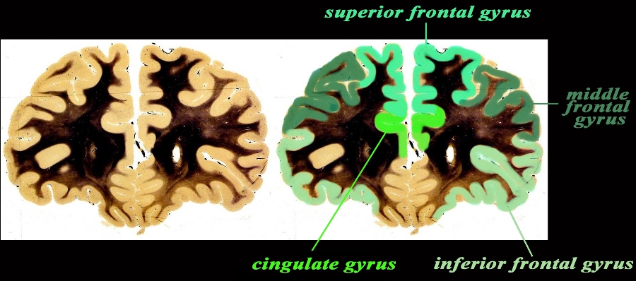
The coronal anatomical image is taken through the frontal lobe just at the extreme anterior portion of the frontal horns. The specimen shows the approximate position of the 3 major frontal gyri in the anterior location including the superior, middle and inferior gyrus. The cingulate gyrus is not really part of the forebrain but part of the limbic system. It holds a medial deep and very central position. code brain forebrain gyrus gyri frontal gyri superior frontal gyrus middle frontal gyrus inferior frontal gyrus cingulate gyrus fx anatomy normal neuroanatomy anatomy normal Cingulate Gyrus Gyrus Rectus normal labelled anatomic sections coronal Boston University BU Courtesy Department of Anatomy and Neurobiology at Boston University School of Medicine Dr. Jennifer Luebke , and Dr. Douglas Rosene Overlays Ashley Davidoff MD 97340.C1.gc01L2.9 |

Gyri at the Vertex – T2 Weighted Axial Image |
|
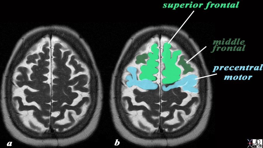
The axial T2 weighted image of a brain that shows significant atrophy as witnessed by widening of the sulci and hence prominence of the gyri of the brain. The axial cut is near the vertex and shows the superior frontal gyrus (bright green) a small portion of the middle frontal gyrus (dark green), and the the precentral gyrus (light blue). The central sulcus lies just posterior to the precentral gyrus. Courtesy Ashley Davidoff MD Copyright 2010 49051c03L.9s |


