The Common Vein Copyright 2010
Definition
Pons means bridge and it is literally a part of the hindbrain that bridges the midbrain and hindbrain. It is part of the brainstem.
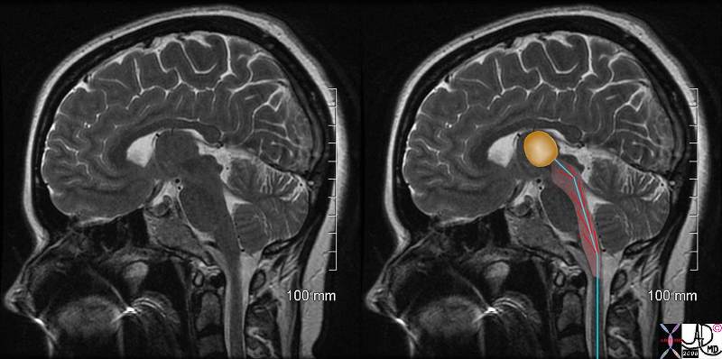
Structurally it is characterised by its position, and its shape. In the coronal projection its anterior component is oval shaped and the posterior portion is more rectangular. The pons is also recognized by the company it keeps. It is closely associated with the fourth ventricle which lies posterior to it. Posterior to the 4th ventricle is the cerebellum. It is approximately 2 cm in diameter and 2.5 cms high.
The pons functions by participating in the transmission of motor and sensory messages to and from the body and face. It is involved with autonomic functions such as respiration, swallowing and bladder function as well as in the function of sleep. It transmits sensory fibers to the thalamus and motor fibers from the forebrain to the cerebellum and medulla.
Diseases of the pons are relatively uncommon but include pontine hemorrhage which is clinically characterized by bilateral pinpoint pupils and which has a mortality of 75% in 24 hours. There is a rare disease called central pontine myelinosis, a demyelinating disease that results in sensory disturbances that include aberrance in touch, motor dysfunction of swallowing and walking and loss of balance.
The Pons in Context The Pons(red) Part of the Hindbrain(Rhombencephalon) Member of the Metencephalon |
|
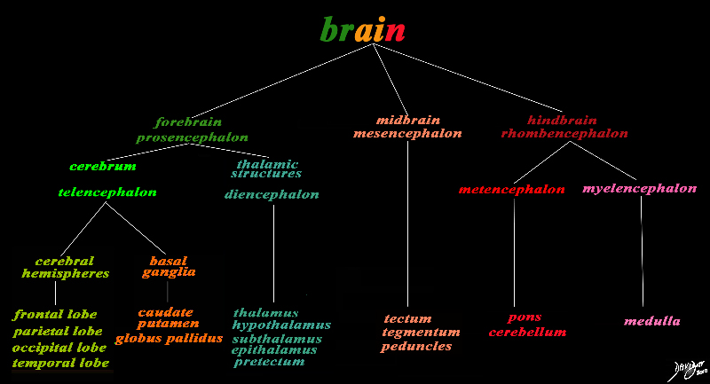
The basic and simplest classification of the brain into forebrain midbrain and hindbrain is shown in this diagram and advanced to a more complex tree using the embryological and evolutionary terminologies. The pons is part of the hindbrain The forebrain consists of the cerebrum also called the prosencephalon, which contains the more advanced form of the brain and the thalamic structures which contain more basic structures. The cerebrum (telencephalon) itself consists of two cerebral hemispheres and paired basal ganglial structures. Each cerebral hemisphere will have gray and white matter distributed in the frontal parietal temporal and occipital lobe, with the basal ganglia being part of the gray matter deep in the cerebral hemispheres. The most important thalamic structures arising from the diencephalons include the thalamus itself and the hypothalamus. The midbrain (mesencepaholon) consists of the tectum tegmentum and cerebral peduncles. The hindbrain has two major branch points based on the evolutionary development. The pons and cerebellum(part of the metencephalon) are grouped and the medulla (part of the myelencephalon is the second branch. Courtesy Ashley Davidoff MD Copyright 2010 All rights reserved 97686.8s |
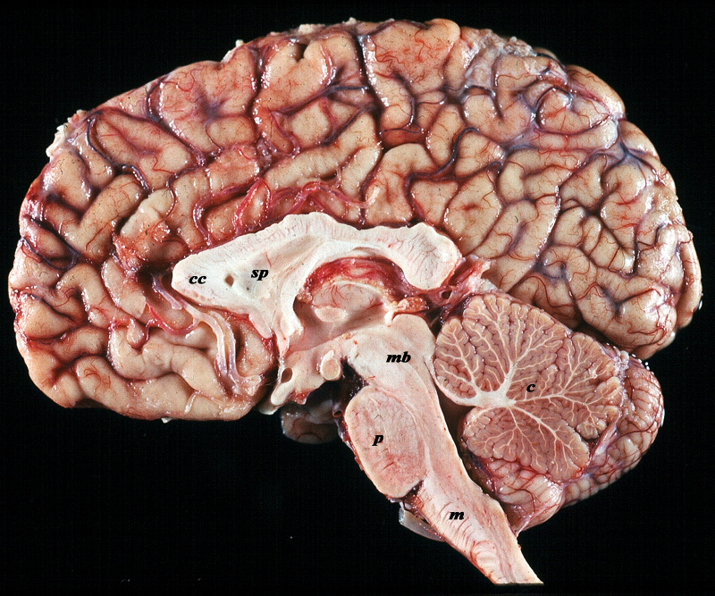
Pons Anatomic Specimen in Midsagittal Section |
|
The midsagittal section view of brain reveals the distinctive shape position and character of the midline structures of the brain. The distinction between the character of the cerebral cortex which has a creamy color and the white matter exemplified by the corpus callosum (c) and septum pellucidum (sp) which are white, and the midbrain (mb) pons (p) and medulla (m) which are off white as opposed to the color of the cerebellum (c) which is light salmon pink is well demonstrated. The relative sizes of the forebrain, midbrain and hindbrain and their components are well appreciated in this section. Image Courtesy of Thomas W.Smith, MD; Department of Pathology, University of Massachusetts Medical School. 97805b02 |
Sagittal Plane
|
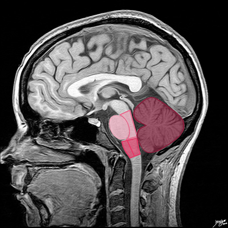
Basic Components of the Pons as seen in the in the Sagittal Plane |
|
The shapes in fact are not quite ovoid. The pons (lightest pink) has shapes reminiscent of an oversize egg lying in a small bed The anterior eggshaped portion is called the ventral or anterior pons and the posterior portion called the tegmentum which is a continuation of the tegmentum of the midbrain. The medulla oblongata is almost rectangular but has a subtle forward bulging to its anterior surface. The anterior portion is called the ventral or anterior medulla and the posterior portion is called the tegmentum, similar in name and a continuation of the tegmentum of the midbrain and pons. The cerebellum is situated posteriorly, is the largest of the three major components and it consists of two hemispheres and a central vermis better appreciated in the axial plane. Courtesy Ashley Davidoff MD Copyright 2010 all rights reserved 92141.3kd03b04b.8s |
Coronal Plane
|
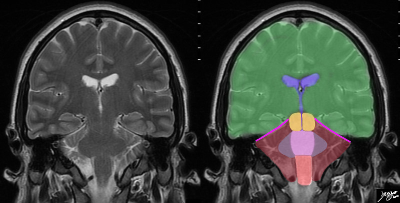
The Pons in Relation to the Medulla Cerebellum and Midbrain |
|
In this T2 weighted coronal MRI image the distinction between the supratentorial and infratentorial structures is made apparent by the bright pink tentorium that acts as a roof of the posterior fossa. The forebrain (green) midbrain (orange) and hindbrain (pink salmon and maroon) and the cerebellum (maroon), with other parts of the hindbrain filled in including the pons (light pink) middle cerebellar peduncles (mauve) and medulla (salmon) All the structures above the pink line are supratentorial, and those below are infratentorial. Part of the midbrain is supratentorial and part is infratentorial. The ventricular system is outlined in blue Courtesy Ashley Davidoff MD copyright 2010 all rights reserved 89721c06b.8sg01 |
|
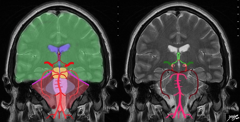
The Vertebrobasilar System and the Pons |
|
The diagram shows the main branches of the blood supply to the brain including the circle of Willis overlaid on coronal MRI image to portray the approximate position of the vessels in the brain. The image on the right shows the combined system all in red, and the image on the right shows the derivation from the vertebrobasilar and carotid systems The carotid system supplies the brain from the internal carotid (salmon pink). We demonstrate its terminal bifurcation into middle cerebral (dark green) and anterior cerebral (bright green). The anterior communicating artery runs between the two anterior cerebrals (bright red) The basilar artery (pink) is formed by the two vertebral arteries and it travels as a single artery over the upper medulla and the pons. Its terminal branch is the posterior cerebral artery (maroon). The first branch off the posterior cerebrals is the posterior communicating which joins the middle cerebral to complete the circle of Willis Each of the carotid and vertebro-basilar systems contributes to the circle of Willis through communicating arteries. The vertebro-basilar system provides the posterior communicating arteries bilaterally from the posterior cerebral and the carotid system provides the anterior communicating arteries via the anterior cerebral arteries. Courtesy Ashley Davidoff MD Copyright 2010 All rights reserved 89721c06b.8sg05.8s |
Axila Plane
|
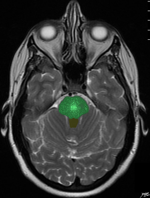
The Pons |
|
The pons is difficult to recognise by intrinsic characteristics, but if you paint it green – render its matrix and paint the 4th ventricle brown it looks like a little bush in a flower pot – non descript in the axial plane – yes! Courtesy Ashley Davidoff copyright 2010 all rights reserved 94084.3kb08.81s |
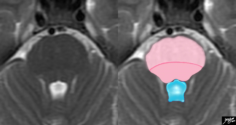
Characteristic 4th Ventricle |
|
TheT2 weighted MRI is a magnified view of the pons recognized commonly in the axial projection by its relationship to the 4th ventricle posteriorly which has a characteristic shape looking like a puffed up crown (blue) The pons consists of an anterior portion called the ventral pons (light pink) and the dorsal portion called the tegmentum ((darker pink). The line of distinction is vague and the dark pink line is drawn in for clarity. Courtesy Ashley Davidoff MD copyright 2010 all rights reserved 94084.3kdc.82sd01 |
|
The Pons and the RAS |
|
The reticular activating system (aka ascending reticular activating system) is a part of the brainconsidered to be the center of arousal and motivation. Structurally it lies betweent the medulla oblongata and midbrain and is connected to the thalamus. In turn al parts of the brain can be stimulated as a result including the cerbral cortex basal regions of the brain and the medulla Functionally it indirectly relates to our state of conciousness, and is involved with the control of the circadian rhythm, respiration, cardiac rhythms, and sexual function. In the instance of painthe RAS is activated by the C fibers and hence pain can arouse us from sleep through the RAS, can create a sense of urgency, and can cause changes in heart rate or respiration rate. Courtesy Ashley Davidoff MD copyright 2008 77059c01b01.8s |
Applied Anatomy Diseases
|
Acute Infarction in the Pons – Vasculitis |
|
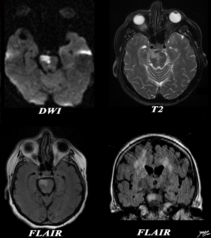
The diffusion weighted MRI is from a 62 year old female patient is with acute neurological deficit The DWI sequence in image (a) reveals a focus of restricted diffusion in the pons. This finding is confirmed on the T 2weighted image (b) as well as on FLAIR on the axial FLAIR (c) and coronal FLAIR(d). On the last image – the coronal image shows FLAIR hyperintensity dominant in the right white matter particularly in the corona radiate. These findings are consistent with an acute infarction in the pons and the multicentricty is compatible with the diagnosis of vasculitis. Image Ashley Davidoff MD Copyright 2010 89038c.8s |
|
DWI – Multicentric Infarction – Acute Vasculitis |
|
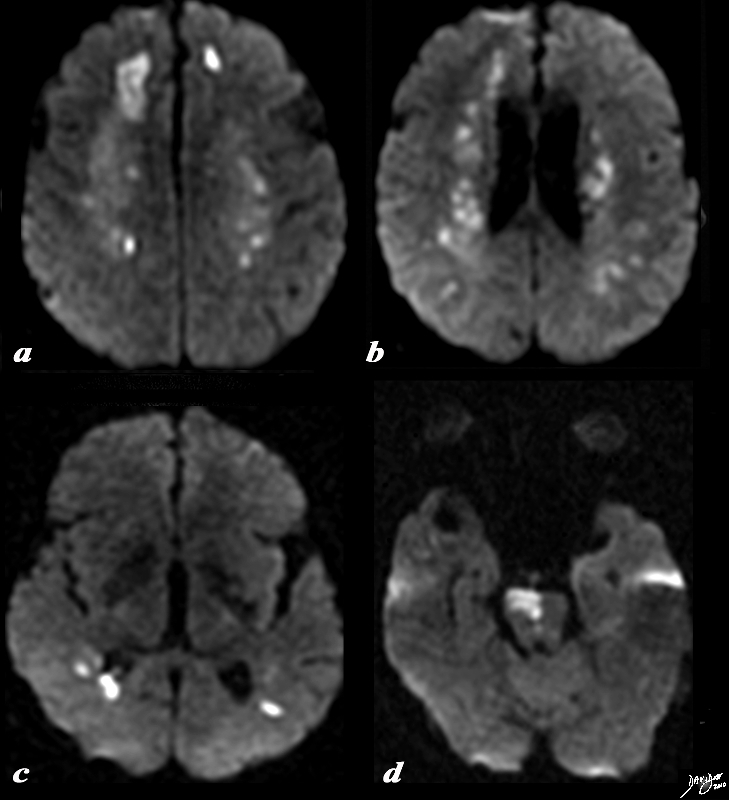
The diffusion weighted MRI is from a 62 year old female patient is with acute neurological deficit The DWI sequence reveals multicentric of restricted diffusion indicating acute infarction. In image a, the dominant infarction is in the right frontal lobe, but there is an area of infarction in the left frontal lobe as well. Image b shows restricted diffusion in the periventricular white matter, while (c) shows disease in the posterior parietal region. In image (d) an infarction of the right side of the pons is apparent. Courtesy Ashley Davidoff MD Copyright 2010 89038c01.8s |
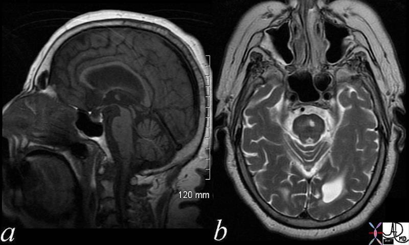
Lacune in the Pons |
|
The MRI is from a 82 year female. In image (a) a vague low density region is seen in the superior as[ect of the pons and on the axial T2 weighted image the chronic infarct is seen as a hyperintense focus. These findings are consistent with a diagnosis of a lacunar infarction in the pons, implying that it is of a chronic nature 71472c02 Image Courtesy Ashley Davidoff MD Copyright 2010 |

