The Common Vein Copyright 2010
Introduction
This section is designed to frame the midbrain to enable basic concepts to start at ground level. It is easiest to conceive the forebrain in three views;- The sagittal, axial and coronal view.
Sagittal
|
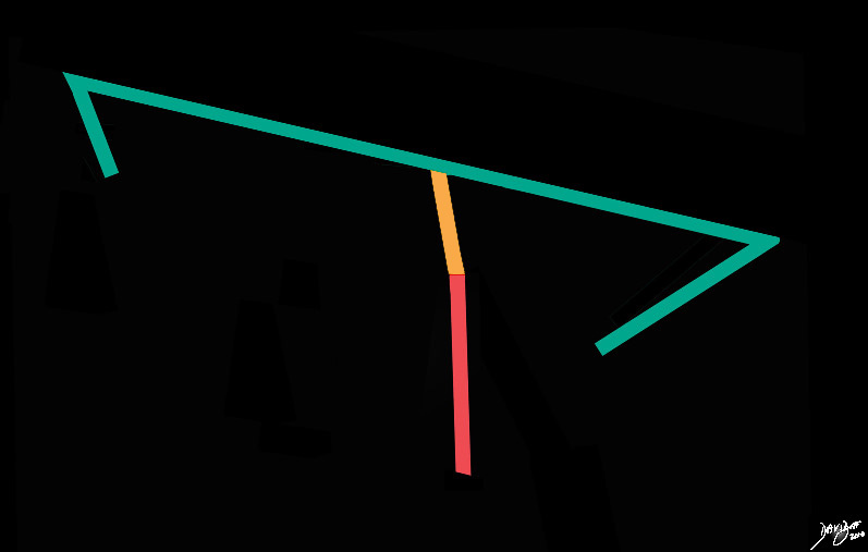
The Midbrain (orange) – A Bridge from the Forebrain (green) to the Hindbrain (salmon) |
|
The stick diagram starts to become more complex as the forebrain (green) demonstrates folds on its anterior and posterior extremes representing the fold of the frontal lobe anteriorly and temporal lobe posteriorly. The vertical limb also has a subtle angulation between the midbrain (yellow) and hind brain (salmon red). Davidoff art Courtesy Ashley Davidoff MD copyright 2010 all rights reserved 93887b03a.81s |
Axial
|
Concepts in the Axila Plane – Two Rectangles Two Ovals and a Tiny Sphere |
|
The midbrain consists conceptually of twoanteroleaterally positioned rectancles, and two posterocentrally placed ovals (purple), surrounding the small aqueduct of Sylvius (white). Courtesy Ashley Davidoff MD copyright 2010 all rights reserved 94074b09b01a0910.8s |
Coronal
|
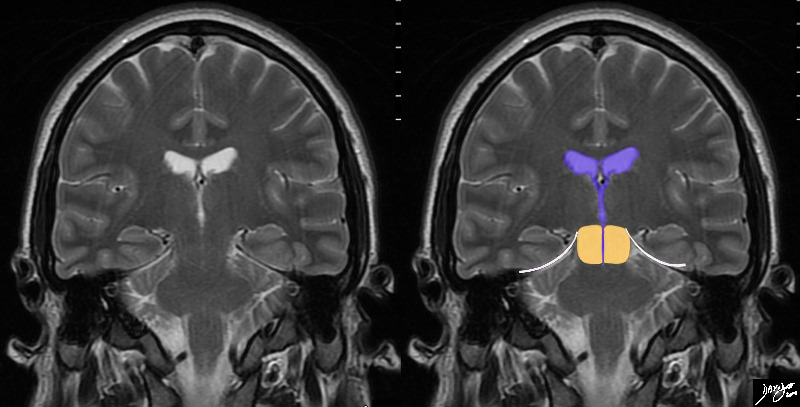
Midbrain in Coronal – Skirting the Tentorium Bridge Between the Forebrain and Hindbrain |
|
In this T2 weighted MRI image – the midbrain (orange) is seen centered aroubnd the ventriclar system. The aqueduct of Sylvius is the finest of channels that courses through the middle of the midbrain, and frequently is the key identifying feature of the midbrain. The temporal lobes rest of the tentorium (white curvilinear convex lines). Below the tenntorium is the midbrain and hind brain. Courtesy Ashley Davidoff MD copyright 2010 all rights reserved 89721c03b01b.8s |


