All posts by adavidoff

Temporal Gyri

Temporal Gyri

The Common Vein Copyright 2010

Introduction

The lateral surface of the temporal lobe is composed of the superior, middle, and inferior temporal gyri.

The superior temporal gyrus continues into the lateral sulcus.

Here it forms one of its walls.

Part of the superior temporal gyrus forms the temporal operculum.

The inferior temporal gyrus continues onto the inferior surface of the lobe.

The rest of the inferior surface is made up of the broad and often discontinuous occipitotemporal (fusiform) gyrus and the parahippocampal gyrus.

These are separated from one another by the collateral sulcus.

The occipitotemporal gyrus is partly in the occipital lobe and partly in the temporal lobe.

The parahippocampal gyrus is continuous with the cingulate gyrus around the splenium of the corpus callosum.

This is by the isthmus of the cingulate gyrus.

The anterior end of the parahippocampal gyrus turns backward and forms a medially directed bump called the uncus.

The superior border of the parahippocampal gyrus is the hippocampal sulcus.

Folded into the temporal lobe at the hippocampal sulcus is the hippocampus.

This is part of the limbic system.

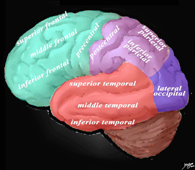
Overview of the Gyri of the Forebrain – Lateral External View

The lateral view of the brain shows the frontal lobe (green) parietal lobe (light mauve), the occipital lobe (purple) and the temporal lobe (red) In this view the frontal lobe gyri that are visible are; superior frontal, middle frontal, inferior frontal and precentral gyri. The parietal gyri include the post central, superior parietal and inferior parietal. The occipital gyrus that is visible is the lateral occipital. The temporal lobe gyri include the superior, middle and inferior temporal gyri.

Courtesy Ashley Davidoff MD Copyright 2010 83029d05g01.8s

Superior Sagittal Sinus

Superior Sagittal Sinus

Ashley Davidoff MD

The Common Vein Copyright 2010

Introduction

Sagittal Sinus


Navy Bluye Part of the C Rings

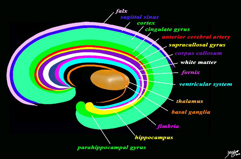
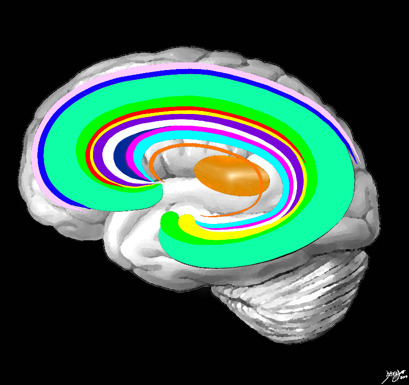
The forebrain has most of its components aligned in a series of inverted c- shaped rings starting from the outer membranes that culminate in the falx (pink), then extending inward smaller inner rings with each intimately connected to the others. The thalamus (dull orange) appears diagrammatically as the centre of these rings as seen from the sagittal view The outer ring is the falx (pink) followed by the sagittal sinus (blue) cerebral cortex (light green), cingulate gyrus (bright green) superiorly which becomes the parahippocampal gyrus inferiorly. The red ring represents the distribution of the main portion of the anterior cerebral artery. Next is the yellow ring which is the supracollosal gyrus (indusium griseum) superiorly and the hippocampus inferiorly. This is followed by the corpus callosum (purple) which enables the white ring of white matter to connect between hemispheres. The next ring is the thin bright pink ring which represents the fornix superiorly and the fimbria inferiorly. The innermost ring (light blue) represents the lateral horns of the ventricular system. The basal ganglia run just lateral to the lateral ventricles. The navy blue arrow headed structure is the septum pellucidum.

Courtesy Ashley Davidoff copyright 2010 all rights reserved 93907d13g03.8s

Foramen of Magendie

Foramen of Magendie aka Median Aperture

Ashley Davidoff MD

The Common vein Copyright 2010

Definition

The foramen of Magendie (aka median aperture) is a single midline structure within the ventricular system of the brain which connects the fourth ventricle with the cerebellopontine cistern. It  represents one of the three pathways that enable CSF to connect with the subarachnoid space.  It lies posterior to the pons and anterior to the cerebellum.

Sagittal View of the Ventricles

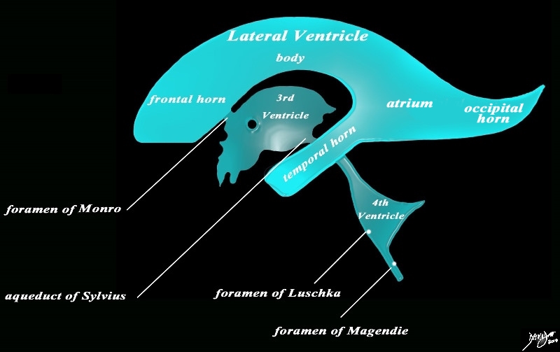
The diagram in the sagittal projection reveals the horizontal portion called the lateral ventricle. It is a paired structure that houses the frontal horn, body, and the vertical portion which is composed of the 3rd ventricle, cerebral aqueduct and the 4th ventricle. The lateral ventricle consists of the frontal horn, body, occipital horn, atrium and the temporal horn The foramen of Monro connects the lateral ventricles with the third ventricle. The paired foramina of Luschka are sitiuated anteriorly in the 4th ventricle and they allow CSF to circulate in the subarachnoid spaces. The foramen of Magendie is a single structure and is situated posteriorly and it also enables CSF to enter the subarachnoid space.

Courtesy Ashley Davidoff MD copyright 2010 all rights reserved 94459b10b02.82s

Temporal  Horns of Ventricles

Temporal  Horns of Ventricles

The Common vein Copyright 2009

Author

Definition

The temporal horns are paired structures of the ventricular system extending from the atria of the ventricles to the temporal lobes.  They course anteriorly lateral ward and then inferiorly extending within 2.5cms of the tip of the temporal lobes.  IKts floor is provided by the hippocampus.

 In a normal adult they are not usually visualized, and if they are  visualised it is may represent the earliest sign of hydrocephalus or cerebral atrophy.


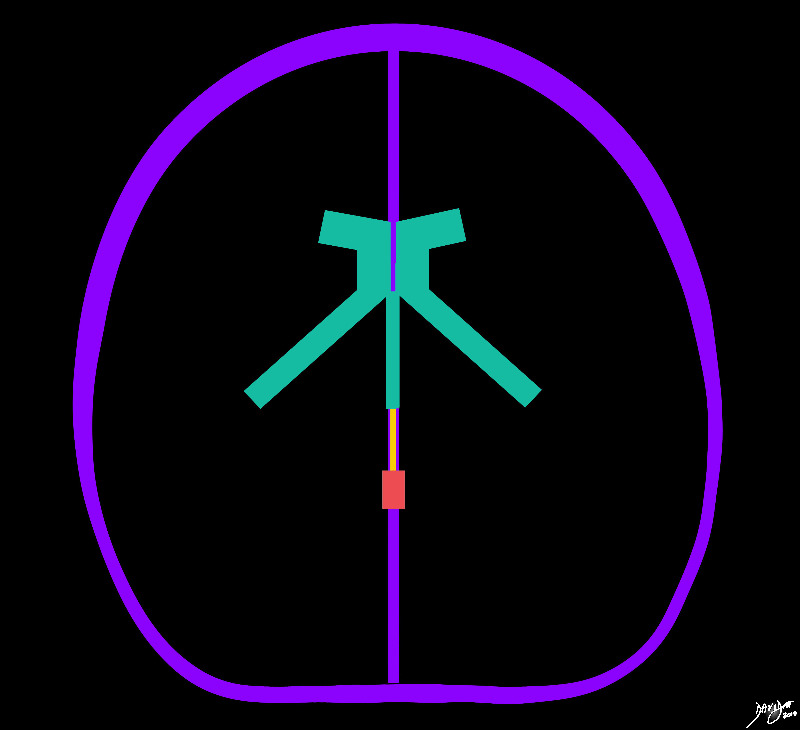
The vectors of the ventricular System Overlaid on the Brain

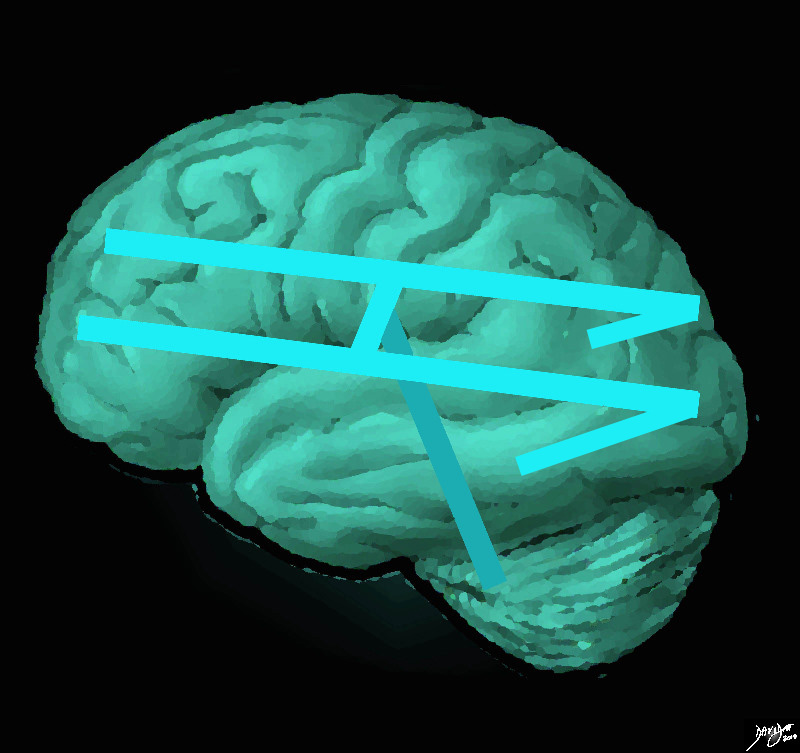
The ventricular system is overlaid on a drawing of the brain. Each limb of the horizontal component is situated in a cerebral hemisphere.  The angled parts that extend inferiorly represent the temporal horns. The vertical limb includes the third and fourth ventricles

Courtesy Ashley Davidoff MD copyright 2010 all rights reserved  94458e07.81s

Sagittal View of the Ventricles

The diagram in the sagittal projection reveals the horizontal portion called the lateral ventricle. It is a paired structure that houses the frontal horn, body, and the vertical portion which is composed of the 3rd ventricle, cerebral aqueduct and the 4th ventricle. The lateral ventricle consists of the frontal horn, body, occipital horn, atrium and the temporal horn The foramen of Monro connects the lateral ventricles with the third ventricle. The paired foramina of Luschka are sitiuated anteriorly in the 4th ventricle and they allow CSF to circulate in the subarachnoid spaces. The foramen of Magendie is a single structure and is situated posteriorly and it also enables CSF to enter the subarachnoid space.

Courtesy Ashley Davidoff MD copyright 2010 all rights reserved 94459b10b02.82s


The Recesses

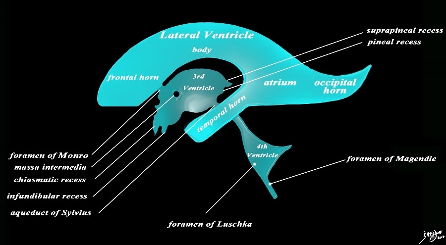
The diagram in the sagittal projection reveals the horizontal portion called the lateral ventricle. It is a paired structure that houses the frontal horn, body, and the vertical portion which is composed of the 3rd ventricle, cerebral aqueduct and the 4th ventricle. The lateral ventricle consists of the frontal horn, body, occipital horn, atrium and the temporal horn The foramen of Monro connects the lateral ventricles with the third ventricle. The paired foramina of Luschka are sitiuated anteriorly in the 4th ventricle and they allow CSF to circulate in the subarachnoid spaces. The foramen of Magendie is a single structure and is situated posteriorly and it also enables CSF to enter the subarachnoid space. code brain ventricles lateral ventricles frontal horn body occipital horn atrium temporal horn 3rd ventricle 4th ventricle foramen of Monro formamen of Magendie Foramen of Luschka anatomy normal neuroanatomy diagram conceptual diagram structure principles Davidoff Art Courtesy Ashley Davidoff MD copyright 2010 all rights

Courtesy Ashley Davidoff MD copyright 2010 all rights reserved  94459b10b02.82s

Temporal Horns – Part of the Forebrain

Distribution of the Ventricles

The distribution of the ventricular system is demonstrated in this diagram with the forebrain structures (green) including the lateral ventriclesfrontal horns, atria, temporal horns, and third ventricle. The midbrain positioned aqueduct of Sylvius is seen in orange, while the hindbrained positioned 4th ventricle is overlaid in orange

Courtesy Ashley DAvidoff MD copyright 2010 all rights reserved 93914b04d07.8s

Temporal Horns Extending from the Atria

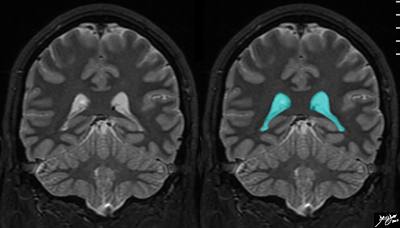
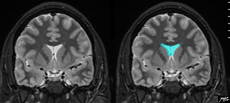
The atria and temporal horns are demonstrated in this coronal MRI using a STIR sequence. The tmporal horns are easily recognized as an inveted ‘V shaped structure starting roughly in the midline and courses laterally and inferiorly. They start just anterior to the occipital lobes and end in the temporal lobes and these lobes can be recognized as neighbours of this part of the ventricular system.

Courtesy Ashley Davidoff MD copyright 2010 all rights reserved 89744c01.8s

Choroid Plexus in the Temporal Horns

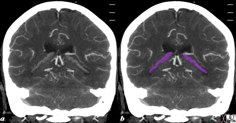
The reconstructed CTA scan in the coronal plane reveals the choroid plexus (purple) as fine knotty strands extending from the atrium to the temporal horns bilaterally.

Image Courtesy Ashley Davidoff MD Copyright 2010 76532c.8s

Applied Biology

Age related Atrophy

Temporal Horns become Visible (d)

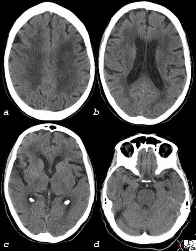
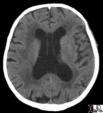
The CT scan of this 92 year old man reveals normal involutional change of the brain including perivntricular lucency(a)suggestive of microangiopathic change, mild dilatation of the ventricles (b) with deepening of the sulci and prominence of the gyri (abc) and ability to identify the temporal horns (d), all signs of brain atrophy.  Note also the normal calcification of the choroid plexus in the atria (c) which is found very commonly even in young patients.

Image Courtesy Ashley Davidoff MD 75932c01

Aqueductal Stenosis

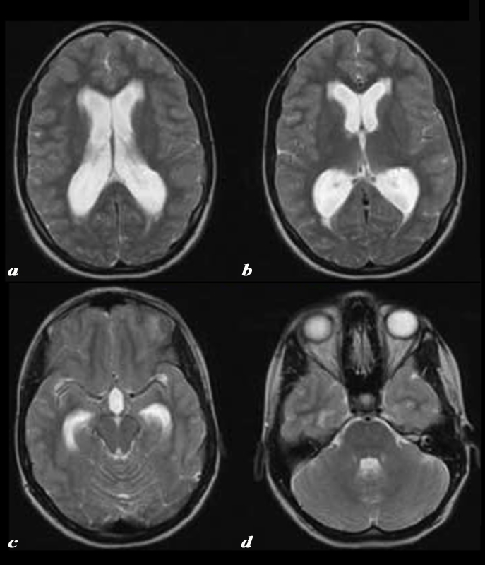
The T2 weighted MRI images show dilated lateral ventricles (a), frontal horns and occipital horns (b), temporal horns (c) and 4th ventricle (d) consistent with the known diagnosis of aqueductal stenosis CODE brain lateral ventricles frontal horns occipital horns atria body of temporal horns 4th ventricle aqueduct of Sylvius fx dilated enlarged distended dx hydrocephalus aqueductal stenosis

Courtesy Philips Medical Systems 92408b.8

Somatosensory Cortex

Somatosensory Cortex

Ashley Davidoff MD

Copyright 2010

Somatosensory Cortex in the Parietal Lobe

The somatosensory cortex is part of the somatosensory system and is characterized by its parietal location in the brain and by its ability to perceive and localize the pain.

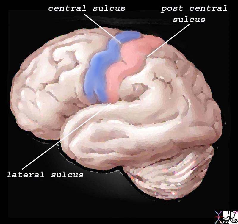
Structurally, the cortex lies as the anterior most structure of the parietal lobe, positioned between between the motor cortex of the frontal lobe and the central sulcus anteriorly, the post central sulcus posteriorly, and the lateral sulcus inferiorly.
The Somatosensory Cortex – Post Central Gyrus

The somatosensory cortex is overlaid in light rose pink in the diagram above and represents the most anterior structure of the parietal lobe.  It lies posterior to the motor cortex (blue) which is part of the frontal lobe, behind the central sulcus and in front of the post central sulcus.  It serves to perceive, localize and evaluate intensity of the pain, as well as initiate the response to the pain.

 83029b01.b1.81s pink = somatosensory cortex in post central gyrus blue = motor cortex The Common vein Davidoff art copyright 2008

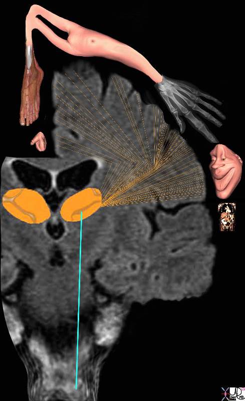
The regions of the body have a specific location in the somatosensory cortex and, depending on the number of nociceptors, will have a a correlatively sized distribution. Thus for example, the lips, mouth, hands, feet, and genitalia will have a much larger representation in the somatosensory cortex than the limbs, trunk, and viscera. Subsequently, the various structures have a descending order of reperesentation and consequently a descending order of sensitivity. The homunculus figure represents this concept and is diagrammed below.

If the somatosensory cortex is viewed in the coronal plane, the homunculus (literally “little man”) is draped over the sensory cortex with genitalia, and legs draped medially, thighs and trunk superiorly, and hands, head, mouth, lips, pharynx, tongue and viscera draped laterally. The size of organ representation is not only specific to pain fibres but related to the sensory and motor system as a whole.
Somatosensory Cortex in the Parietal Lobe

Localization and the Homunculus Man

The diagram reflects the relative functional sensory space each body part occupies in the somatosensory cortex.  Those structures with  high density of sensory receptors are represented by a larger size, while those with a lesser concentration of sensory apparatus shown as being “smaller” in size. Hence the mouth lips, hands feet and genitalia have a relatively large representation. The homunculus man (literally the “little man”) is the distorted figure drawn to reflect the concept of size of organ paralleling the size of the sensory innervation.

somatosensory cortex (sensory homunculus) spinothalamic tract spinal cord thalamus sensory cortex homunculus man penis clitoris genitals genitalia foot body thigh abdomen chest and face mouth eyes lips viscera somatosensory Davidoff art Copyright 2008 38610b09.46k.8s

The function of the somatosensory cortex is that of a higher processing centre for touch, temperature, pain, and proprioception serving to amplify awareness of the sensations enabled by the thalamus. Sensation from the left side of the body are processed in the right somatosensory cortex and similarly those from the right side are processed on the left. The higher function of the somatosensory cortex allows us to localise the pain to a specific site, perceive the character and intensity of the stimulus, and sometimes helps identify the shape of the originating object.

The somatosensory cortex is not the final level of the somatosensory system since it also relays impulses to other cerebral areas of perception and reaction. Thus it sends signals via the white matter to other centres in the cortex to enable integration with visual and auditory input, and with other higher cortical functions such as emotion and memory for example. The full experience is then “seen” by the brain enabling the consequent reaction to be as discriminating  and prudent as the nature and experience of the person allows. The difference between the reaction of an infant, child and an adult to the “shot at the doctors” speaks volumes about this latter function.
Septum Pellucidum

Septum Pellucidum

Ashley Davidoff MD

The Common Vein Copyright 2010

Definition

The septum pellucidum is a thin traingular membrane that partitions the frontal horn of the lateral ventricles. It runs vertically between the corpus callosum superiorly and the rostrum of the corpus callosum and the fornix inferiorly.

Structurally it consists of two layers that contain gray and white matter which are usually fused, but may be separated into two as a normal variant.  This condition is called cavum septum pellucidum.

Functionally it participated in emotions of pleasure and rage, mood and sexual gratification.

Disease in the septum pellucidum results in cognitive impairment, as well as vision and coordination problems.

Diagnosis is by MRI and treatment is supportive.

Septum Pellucidum

Part of the Inverted C Rings – Navy Blue

The forebrain has most of its components aligned in a series of inverted c- shaped rings starting from the outer membranes that culminate in the falx (pink), then extending inward smaller inner rings with each intimately connected to the others. The thalamus (dull orange) appears diagrammatically as the centre of these rings as seen from the sagittal view The outer ring is the falx (pink) followed by the sagittal sinus (blue) cerebral cortex (light green), cingulate gyrus (bright green) superiorly which becomes the parahippocampal gyrus inferiorly. The red ring represents the distribution of the main portion of the anterior cerebral artery. Next is the yellow ring which is the supracollosal gyrus (indusium griseum) superiorly and the hippocampus inferiorly. This is followed by the corpus callosum (purple) which enables the white ring of white matter to connect between hemispheres. The next ring is the thin bright pink ring which represents the fornix superiorly and the fimbria inferiorly. The innermost ring (light blue) represents the lateral horns of the ventricular system. The basal ganglia run just lateral to the lateral ventricles. The navy blue arrow headed structure is the septum pellucidum.

Courtesy Ashley Davidoff copyright 2010 all rights reserved 83027b04g03.8s

Septum Pellucidum in between the Frontal Horns 

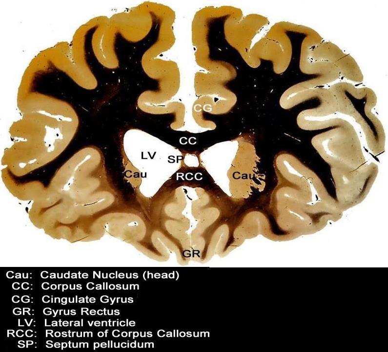
 This coronal section through the forebrain is an anterior cut through the frontal lobes recognized by absence of the temporal horns, the presence of the rostrum of the corpus callosum (RCC), the cingulate gyrus (CG) superiorly and the gyrus rectus (GR) inferiorly. The visualized cortex is therefore part of the frontal lobe, and the ventricles (LV) that are visualized represent the frontal horns. The caudate nuclii (CN) are seen inferolateral to the frontal horns, and the septum pellucidum is seen medially

Courtesy Department of Anatomy and Neurobiology at Boston University School of Medicine Dr. Jennifer Luebke, and Dr. Douglas Rosene 97342.C3.8L01

Gross Anatomy of the Septum Pellucidum

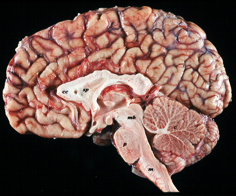
The midsagittal section view of brain reveals the distinctive shape position and character of the midline structures of the brain. The distinction between the character of the cerebral cortex which has a creamy color and the white matter exemplified by the corpus callosum (c) and septum pellucidum (sp) which are white, and the midbrain (mb) pons (p) and medulla (m) which are off white as opposed to the color of the cerebellum (c) which is light salmon pink is well demonstrated. The relative sizes of the forebrain, midbrain and hindbrain and their components are well appreciated in this section.

Image Courtesy of Thomas W.Smith, MD; Department of Pathology, University of Massachusetts Medical School. 97805b02

Septum Pellucidum in Coronal Projection

The frontal horns are seen in this coronal MRI using a STIR sequence. They are easily recognized as a T shaped midline structure, divided in the middle by the gracile septum pellucidum. The frontal horns course through the frontal lobes and above the anterior portion of the temporal lobes. At this level many of the components of the basal ganglia can be evaluated below and lateral to the frontal horns c

Courtesy Ashley Davidoff MD copyright 2010 all rights reserved 89732c01.8s

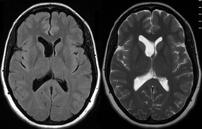
Asymmetry of the Lateral Ventricles – Minor Shift of the Septum Pellucidum – Normal Variant

The MRI shows a FLAIR and a T2 weighted image revealing normal asymmetric development of the lateral ventricles in a 41 year old female.

Courtesy Ashley Davidoff MD Copyright 2010 89044c


Cavum Septum Pellucidum

This CT demonstrates widening of the 2 septal laminae of the septum pellucidum in the lateral ventricles This condition is called cavum septum pellucidum which is a structural deformity of the septum pellucidum where there is a separation between its two leaflets (septal laminae) which have failed to fuse in the first 3-6 months of life. This condition is seen in 100% of fetuses , and about 85% fuse by 3-6 months

Courtesy Ashley Davidoff MD Copyright All rights Reserved 49030

Acute Hemorrhagic Stroke of the Basal Ganglia

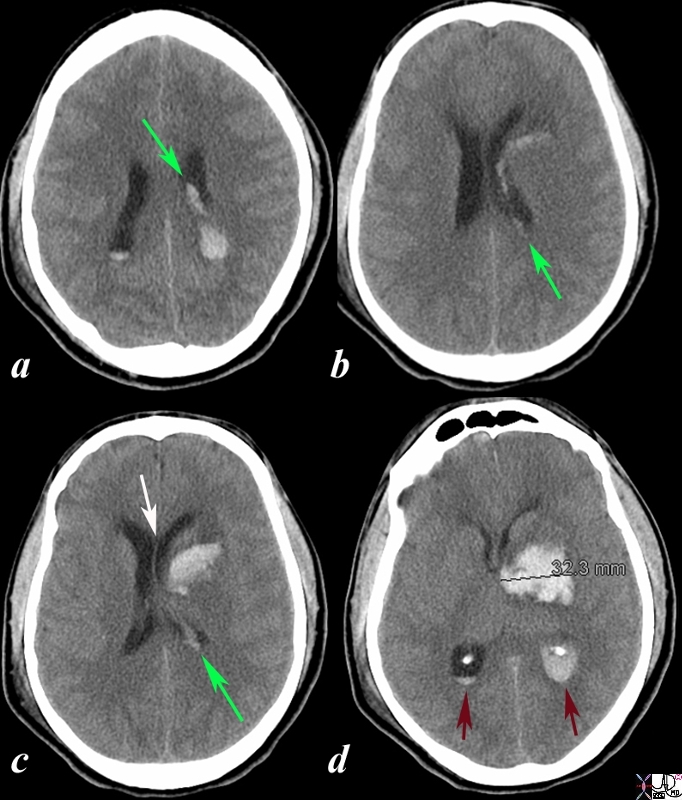
Deviation of Septum Pellucidum

The CT is from a 33year old male with an acute left basal ganglial hemorrhagic stroke. The CT shows a hyperdense accumulation of hemorrhage(d) complicated by extension or rupture of the hemorrhage into the ipsilateral choroid plexus (green arrows a,b,c) and hemorrhage into the ventricles with blood lying dependently in the occipital horns (maroon arrows in c) and midline shift with septum pellucidum (white arrow of the eyes (lenses overlaid in d) mass effect on the left frontal horn (d) and midline shift exemplified by the shift of the septum pellucidum (white arrow c).

Image Courtesy Ashley Davidoff MD Copyright 2010 98551cL.8s

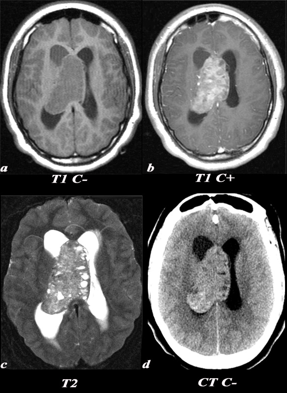
Central Neurocytoma

Extends via the Lateral Ventricle to the Septum Pellucidum

A 21 year old female was found to have papilledema on physical exam noted by her optometrist.

MRI: T1 pre (T1 C- a) and post (T1 C+ b): Post contrast images demonstrate the heterogeneous enhancing nature of this mass. These images demonstrate centered in the right lateral ventricle with involvement of the septum pellucidum and partial extension into the left lateral ventricle. T2: T2 weighted images demonstrate internal areas of high signal consistent with cystic components. Also note the high T2 signal adjacent to the enlarged lateral ventricles which is a finding consistent with hydrocephalus, classically called transependymal flow of CSF. This unenhanced

CT scan demonstrates also demonstrates the mass in the right lateral ventricle with similar features of heterogeneity and involvement of the septum pellucidum and partial extension into the left lateral ventricle. Notice the internal low density or cystic components and the higher density calcifications seen posteriorly. The lateral ventricles are enlarged consistent with resultant hydrocephalus.

The diagnosis is consistent with a central neurocytoma.

Image Courtesy Elisa Flower MD and AsimMian MD 97634c01.8s

Posterior Commissure

Posterior Commissure

The Common Vein Copyright 2009

Definition

The posterior commissure is a rounded band of axons crossing the cerebral aqueduct.

The posterior commissure is responsible for controlling the pupillary response to light.

It is usually observed through MRI. Disease results in abnormal pupillary response. It is also been found to have abnormalities in Parkinson’s disease and Niemann-Pick disease.

Treatments for the alleviation of symptoms and discomfort are in general medical and surgical depending on th disease.

Foramen of Monro

Foramen of Monro

Author

 Copyright 2010

Definition

The foramen of Munro is a narrow and short channel that connects the lateral ventricles with the third ventricle. Each lateral ventricle has a foramen of Munro which leads into a narrow channel called the sulcus of Monro. to join the 3rd ventricle.  They are sitiated just posterior to the frontal horns and anterior to the body of the ventricle.

Structurally they are recognised by their position and small  size.

Functionally they form an important channel to enable CSF to circulate from the larger lateral ventricles to the rest of the ventricular system and subarachnoid space.

Although they themselves are not the origin of disease, they may be easily obstructed by masses or hematomas in the area.  They are there froe sometimes the site of obstructio and manifest with upstream hydrocephalus.

Often their identification by imaging is by inference of their position rather than direct visualization since they lie between the lateral ventricles and the superoanterior position of the 3rd ventricle.

Diagnosis of disease is usually by imaging either by MRI or by CT.

Treatment of hydrocephalus is either directed at the cause or by shunting.


The vectors of the ventricular System Overlaid on the Brain

The ventricular system is overlaid on a drawing of the brain. Each limb of the horizontal component is situated in a cerebral hemisphere.  The angled parts that extend inferiorly represent the temporal horns. The vertical limb includes the third and fourth ventricles

Courtesy Ashley Davidoff MD copyright 2010 all rights reserved  94458e07.81s

Sagittal View of the Ventricles – Basics

The diagram in the sagittal projection reveals the horizontal portion called the lateral ventricle. It is a paired structure that houses the frontal horn, body, and the vertical portion which is composed of the 3rd ventricle, cerebral aqueduct and the 4th ventricle. The lateral ventricle consists of the frontal horn, body, occipital horn, atrium and the temporal horn The foramen of Monro connects the lateral ventricles with the third ventricle. The paired foramina of Luschka are sitiuated anteriorly in the 4th ventricle and they allow CSF to circulate in the subarachnoid spaces. The foramen of Magendie is a single structure and is situated posteriorly and it also enables CSF to enter the subarachnoid space.

Courtesy Ashley Davidoff MD copyright 2010 all rights reserved 94459b10b02.82s


Recesses

The diagram in the sagittal projection reveals the horizontal portion called the lateral ventricle. It is a paired structure that houses the frontal horn, body, and the vertical portion which is composed of the 3rd ventricle, cerebral aqueduct and the 4th ventricle. The lateral ventricle consists of the frontal horn, body, occipital horn, atrium and the temporal horn The foramen of Monro connects the lateral ventricles with the third ventricle. The paired foramina of Luschka are sitiuated anteriorly in the 4th ventricle and they allow CSF to circulate in the subarachnoid spaces. The foramen of Magendie is a single structure and is situated posteriorly and it also enables CSF to enter the subarachnoid space. code brain ventricles lateral ventricles frontal horn body occipital horn atrium temporal horn 3rd ventricle 4th ventricle foramen of Monro formamen of Magendie Foramen of Luschka anatomy normal neuroanatomy diagram conceptual diagram structure principles Davidoff Art Courtesy Ashley Davidoff MD copyright 2010 all rights

Courtesy Ashley Davidoff MD copyright 2010 all rights reserved  94459b10b02.82s

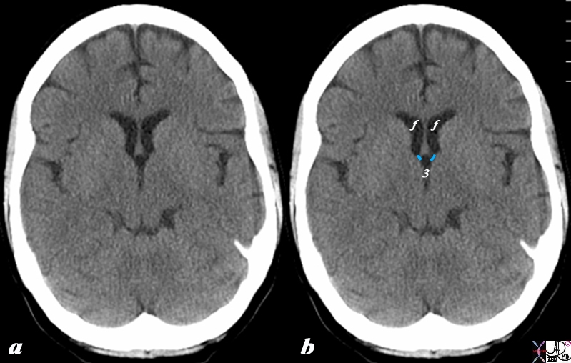
Foramina of Munro Defining the Posterior Border of the Frontal Horns

The CT scan in axial projection reveals the frontal horns (f) connecting with the 3rd ventricle (3) via the small foramina of Munro (overlaid in light blue). The posterior border of the frontal horns is defined by the foramen of Munro on either side.

Courtesy Ashley Davidoff MD Copyright 2010 All rights reserved 98169cL.8s

Normal Asymmetry of the Lateral Ventricles

Symmetry of the Small Foamina of Munro

The MRI shows a FLAIR and a T2 weighted image revealing normal asymmetric development of the lateral ventricles in a 41 year old female.  Note however the two symmetric small channels representing the foramina of Munro which are not affected by the asymmetry.

Courtesy Ashley Davidoff MD Copyright 2010 89044c

Foramen of Magendie

Foramen of Magendie aka Median Aperture

Ashley Davidoff MD

The Common vein Copyright 2010

Definition

The foramen of Magendie (aka median aperture) is a single midline structure within the ventricular system of the brain which connects the fourth ventricle with the cerebellopontine cistern. It  represents one of the three pathways that enable CSF to connect with the subarachnoid space.  It lies posterior to the pons and anterior to the cerebellum.

Sagittal View of the Ventricles

The diagram in the sagittal projection reveals the horizontal portion called the lateral ventricle. It is a paired structure that houses the frontal horn, body, and the vertical portion which is composed of the 3rd ventricle, cerebral aqueduct and the 4th ventricle. The lateral ventricle consists of the frontal horn, body, occipital horn, atrium and the temporal horn The foramen of Monro connects the lateral ventricles with the third ventricle. The paired foramina of Luschka are sitiuated anteriorly in the 4th ventricle and they allow CSF to circulate in the subarachnoid spaces. The foramen of Magendie is a single structure and is situated posteriorly and it also enables CSF to enter the subarachnoid space.

Courtesy Ashley Davidoff MD copyright 2010 all rights reserved 94459b10b02.82s