Frontal Horns of Ventricles
Copyright 2010
Definition
The frontal horns are part of the ventricular system of the brain and more specifically part of the lateral ventricles. They are the most anterior and superior part of the lateral ventricle and are delineated from the body of the lateral ventricles by the foramen of Munro. media;lly they are bounded by the septum pellucidum and inferiorly by the head of the caudate nucleus. The corpus callosum runs superior to them.
|
The vectors of the ventricular System Overlaid on the Brain |
|
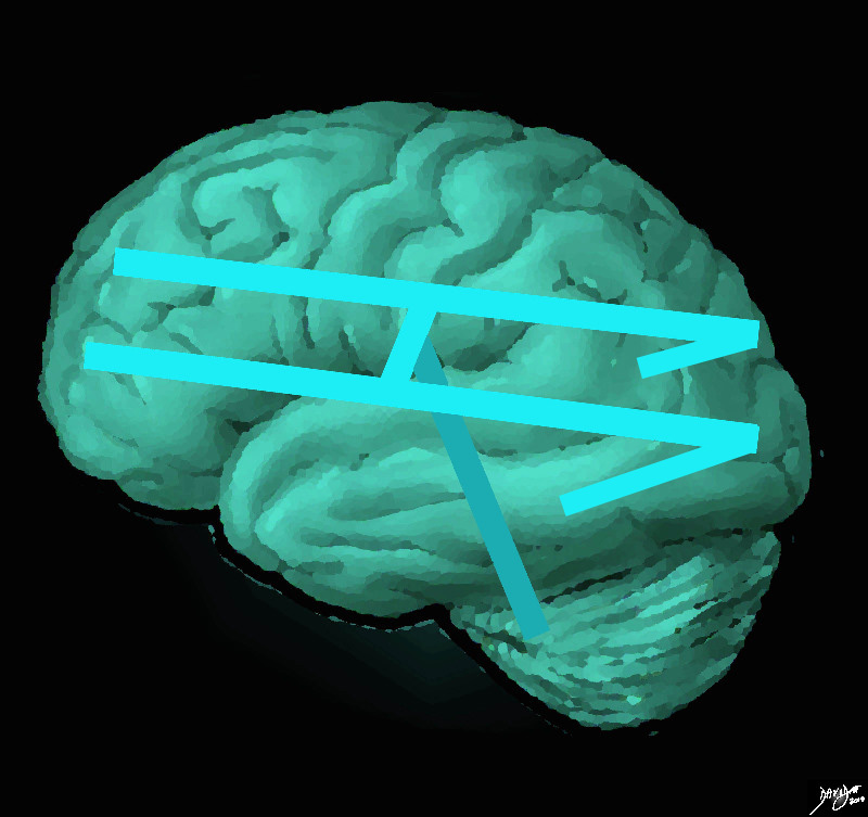
The ventricular system is overlaid on a drawing of the brain. Each limb of the horizontal component is situated in a cerebral hemisphere. The angled parts that extend inferiorly represent the temporal horns. The vertical limb includes the third and fourth ventricles Courtesy Ashley Davidoff MD copyright 2010 all rights reserved 94458e07.81s |
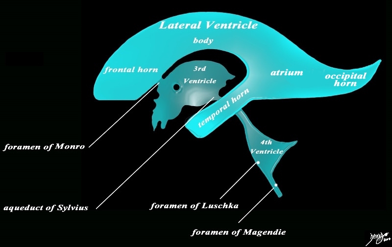
Sagittal View of the Ventricles – Basics |
|
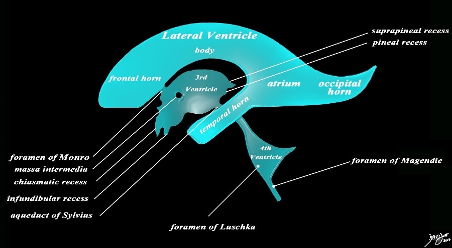
The diagram in the sagittal projection reveals the horizontal portion called the lateral ventricle. It is a paired structure that houses the frontal horn, body, and the vertical portion which is composed of the 3rd ventricle, cerebral aqueduct and the 4th ventricle. The lateral ventricle consists of the frontal horn, body, occipital horn, atrium and the temporal horn The foramen of Monro connects the lateral ventricles with the third ventricle. The paired foramina of Luschka are sitiuated anteriorly in the 4th ventricle and they allow CSF to circulate in the subarachnoid spaces. The foramen of Magendie is a single structure and is situated posteriorly and it also enables CSF to enter the subarachnoid space. Courtesy Ashley Davidoff MD copyright 2010 all rights reserved 94459b10b02.82s |
|
Detailed Components |
|
The diagram in the sagittal projection reveals the horizontal portion called the lateral ventricle. It is a paired structure that houses the frontal horn, body, and the vertical portion which is composed of the 3rd ventricle, cerebral aqueduct and the 4th ventricle. The lateral ventricle consists of the frontal horn, body, occipital horn, atrium and the temporal horn The foramen of Monro connects the lateral ventricles with the third ventricle. The paired foramina of Luschka are sitiuated anteriorly in the 4th ventricle and they allow CSF to circulate in the subarachnoid spaces. The foramen of Magendie is a single structure and is situated posteriorly and it also enables CSF to enter the subarachnoid space. code brain ventricles lateral ventricles frontal horn body occipital horn atrium temporal horn 3rd ventricle 4th ventricle foramen of Monro formamen of Magendie Foramen of Luschka anatomy normal neuroanatomy diagram conceptual diagram structure principles Davidoff Art Courtesy Ashley Davidoff MD copyright 2010 all rights Courtesy Ashley Davidoff MD copyright 2010 all rights reserved 94459b10b02.82s |
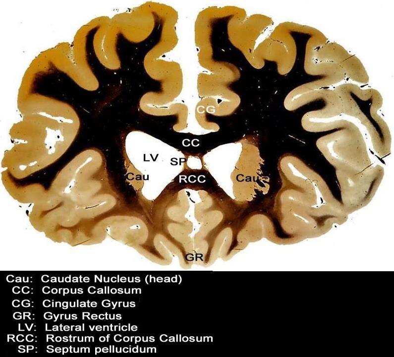
Frontal Horns – Septum Pellucidum Medially,Rostrum of Corpus Callosum Inferiorly and Cingulate Gyrus Superiorly |
|
This coronal section through the forebrain is an anterior cut through the frontal lobes recognized by absence of the temporal horns, the presence of the rostrum of the corpus callosum (RCC), the cingulate gyrus (CG) superiorly and the gyrus rectus (GR) inferiorly. The visualized cortex is therefore part of the frontal lobe, and the ventricles (LV) that are visualized represent the frontal horns. The caudate nuclii (CN) are seen inferolateral to the frontal horns, and the septum pellucidum is seen medially Courtesy Department of Anatomy and Neurobiology at Boston University School of Medicine Dr. Jennifer Luebke, and Dr. Douglas Rosene 97342.C3.8L01 |
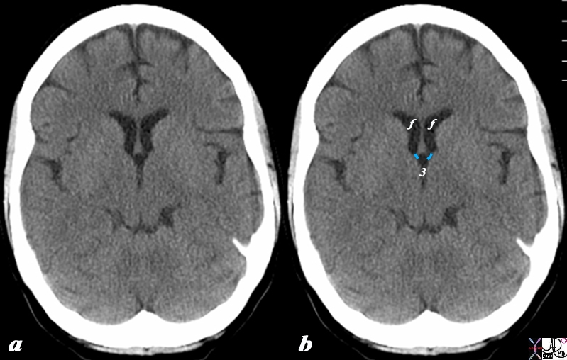
Foramina of Munro Defining the Posterior Border of the Frontal Horns |
|
The CT scan in axial projection reveals the frontal horns (f) connecting with the 3rd ventricle (3) via the small foramina of Munro (overlaid in light blue). The posterior border of the frontal horns is defined by the foramen of Munro on either side. Courtesy Ashley Davidoff MD Copyright 2010 All rights reserved 98169cL.8s |
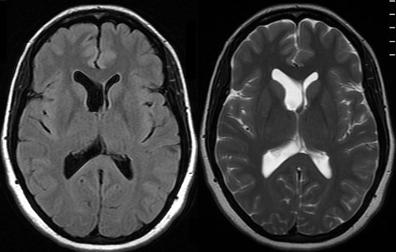
Normal Asymmetry of the Lateral Ventricles |
|
The MRI shows a FLAIR and a T2 weighted image revealing normal asymmetric development of the lateral ventricles in a 41 year old female. Courtesy Ashley Davidoff MD Copyright 2010 89044c |
Applied Biology- Diseases
|
70 year Brain in Two Different Patients “Normal” Involution (a) Normal Pressure Hydrocephalus (b) |
|
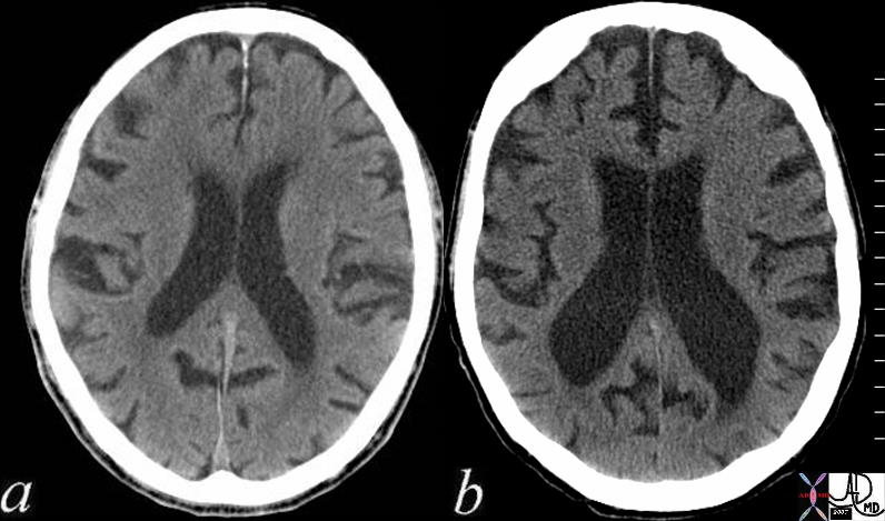
The 2 CT scans show 70 year old brains in two different patients. The patient on the left (a), reveals normal involution of the brain characterized by deepening of the sulci and prominence of the gyri together with a prominent ventricular system. Contrast this brain with the CT scan of the 22 year old above where the sulci and gyri are barely distinguished and the ventricles are slit like. The second patient (b) has a condition called normal pressure hydrocephalus (NPH). In addition to having “normal” involution, the patient also has dilated ventricles more than one would expect for the degree of involution. This is an abnormal finding. Courtesy Ashley Davidoff MD copyright 2010 72179c01 |
Hemorrhage
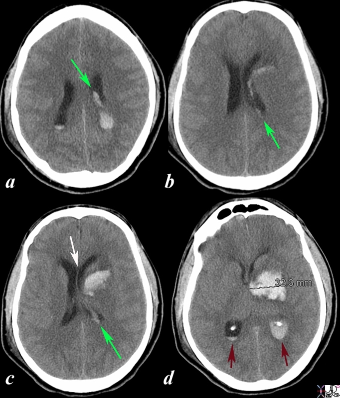
Acute Hemorrhagic Stroke of the Basal Ganglia Mass Effect on the Frontal Horn |
|
The CT is from a 33year old male with an acute left basal ganglial hemorrhagic stroke. The CT shows a hyperdense accumulation of hemorrhage(d) complicated by extension or rupture of the hemorrhage into the ipsilateral choroid plexus (green arrows a,b,c) and hemorrhage into the ventricles with blood lying dependently in the occipital horns (maroon arrows in c) and midline shift with septum pellucidum (white arrow of the eyes (lenses overlaid in d) mass effect on the left frontal horn (d) and midline shift exemplified by the shift of the septum pellucidum (white arrow c). Image Courtesy Ashley Davidoff MD Copyright 2010 98551cL.8s |
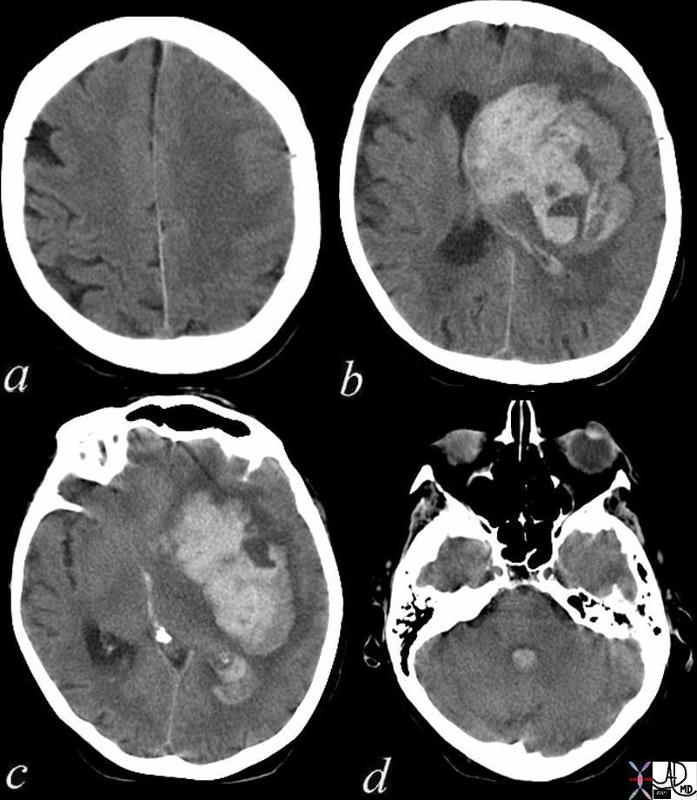
Acute Cerebral Hemorrhage with Mass Effect and Rupture into the Ventricles |
| This CT shows an acute hemorrhagic event originating in the frontoparietal region of the left cerebral hemisphere causing significant mass effect by compressing and displacing the ipsilateral lateral ventricle with significant midline shift.
The hemorrhage has ruptured into the ipsilateral lateral ventricle and blood can be seen within the choroid plexus (b) in the posterior horn (c) as well as the 4th ventricle (d). The ipsilateral edema has caused loss of the gray white matter interface in the left parietal lobe Courtesy Ashley Davidoff MD 72143c01 |
|
Aqueductal Stenosis and Hydrocephalus |
|
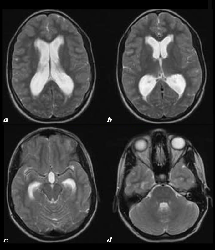
The T2 weighted MRI images show dilated lateral ventricles (a), frontal horns and occipital horns (b), temporal horns (c) and 4th ventricle (d) consistent with the known diagnosis of aqueductal stenosis CODE brain lateral ventricles frontal horns occipital horns atria body of temporal horns 4th ventricle aqueduct of Sylvius fx dilated enlarged distended dx hydrocephalus aqueductal stenosis Courtesy Philips Medical Systems 92408b.8 |

