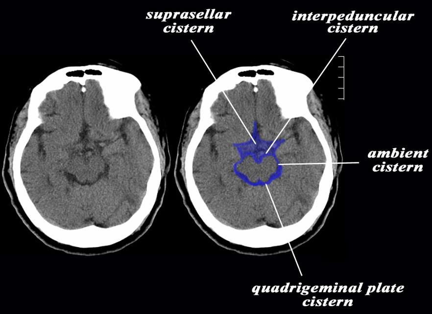
Cisterna Magna
Sumit Karia MD
The Common Vein Copyright 2010
Definition
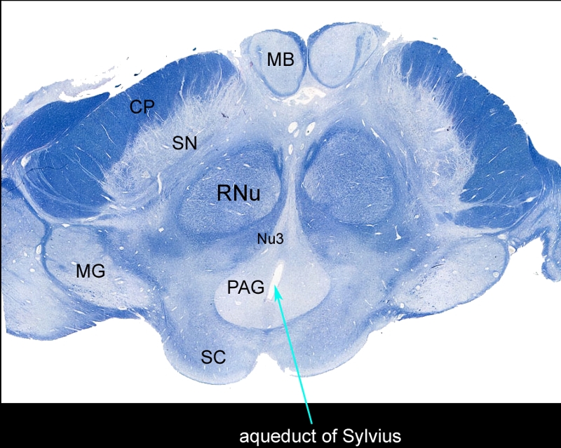
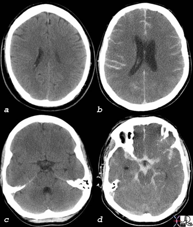
The cisterna magna (or cerebellomedullary cistern) is one of the three principal cisterns. It is located between the cerebellum and the dorsal surface of the medulla oblongata.
Anteriorly it abuts the posterior surface of the medulla
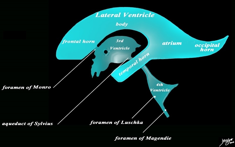
This large cistern communicates with the fourth ventricle through the foramen of Magendie and distally is continuous with the subarachnoid space around the spinal cord.
The cisterna magna contains large vessels including the vertebral artery and posterior inferior cerebellar artery. It also houses the inferior cerebellar tonsils.
In case of intracranial hypertension, these tonsils can migrate to the foramen magnum and compress the medulla located just in front. This constitutes a risk of lumbar punctures.
|
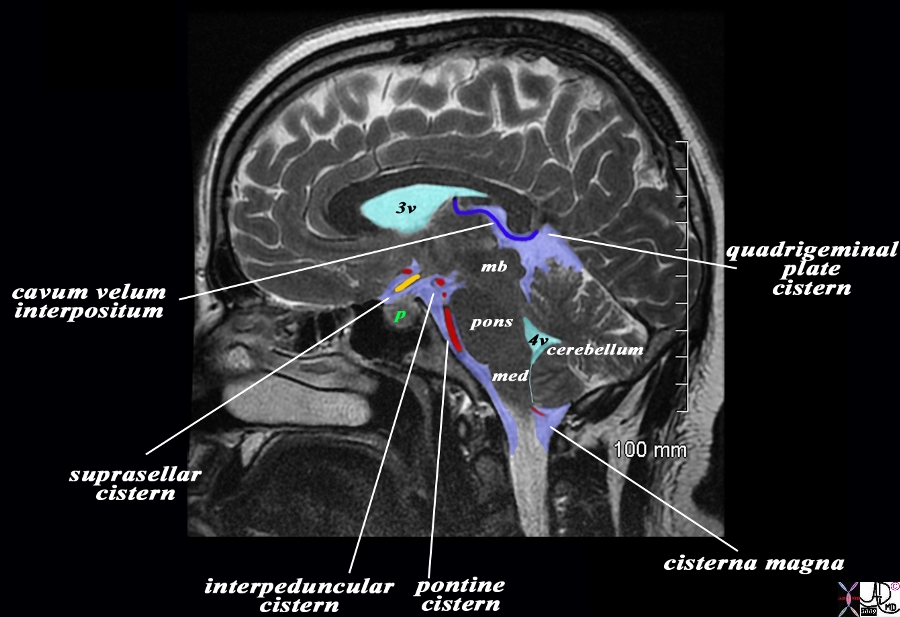
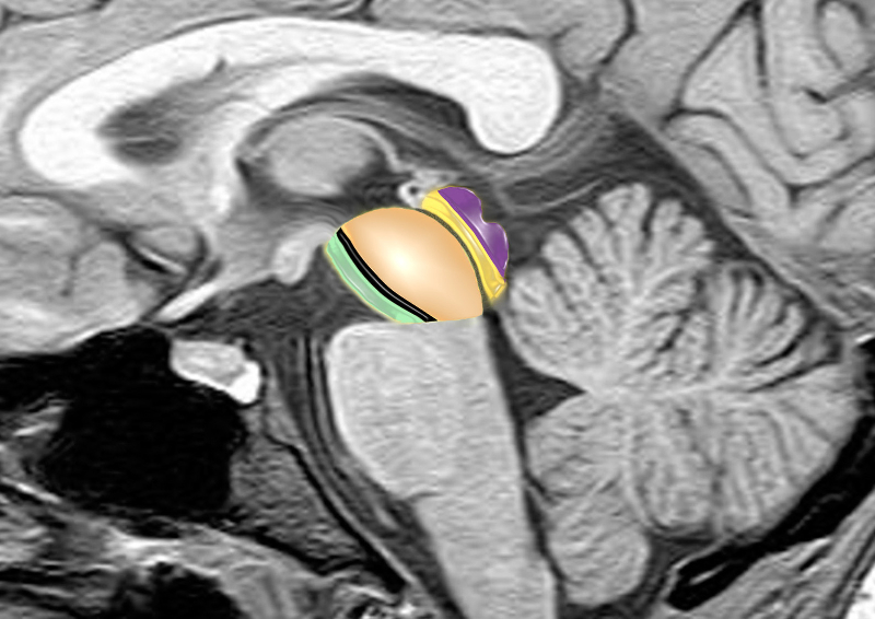
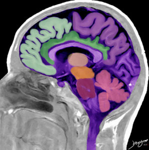
Basal Cisterns in Sagittal Section |
|
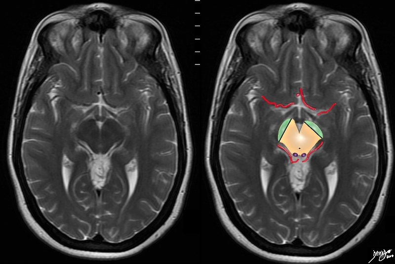
The overlay outlines the basal cisterns in light blue, and the ventricles (teal blue), and a variety of cerebral structures including the midbrain (mb) pons, medulla (med) and cerebellum. A variety of blood vessels (overlaid in blue and red are related to the basal cisterns. The image is T2 weighted sagittal MRI sequence from a patient with a macroadenoma of the pituitary (green p). Starting inferior and anterior we follow the pontine or prepontine cistern associated with the basilar artery, the interpeduncular cistern which is an anterior to the peduncles of the midbrain, the suprasellar cistern (above the pituitary – green p), and the cavum interpositum. Posteriorly the quadrigeminal plate cistern lies behind the colliculi and finally the cisterna magna which surrounds the inferior aspect of the medulla and cerebellum and merges with the subarachnoid space around the cord. The peripheral dural sinuses include the superior sagittal sinus, the laterally placed junction of the transverse sinus and straight sinus, and the confluens of the inferior sagittal sinus which runs on the inferior surface of the falx and the straight sinus which lies on the posterior and superior surface of the tentorium overlying the cerebellum.
Image Courtesy Ashley Davidoff MD Copyright 2010 All rights reserved 98287b03L02b.9s
|
|
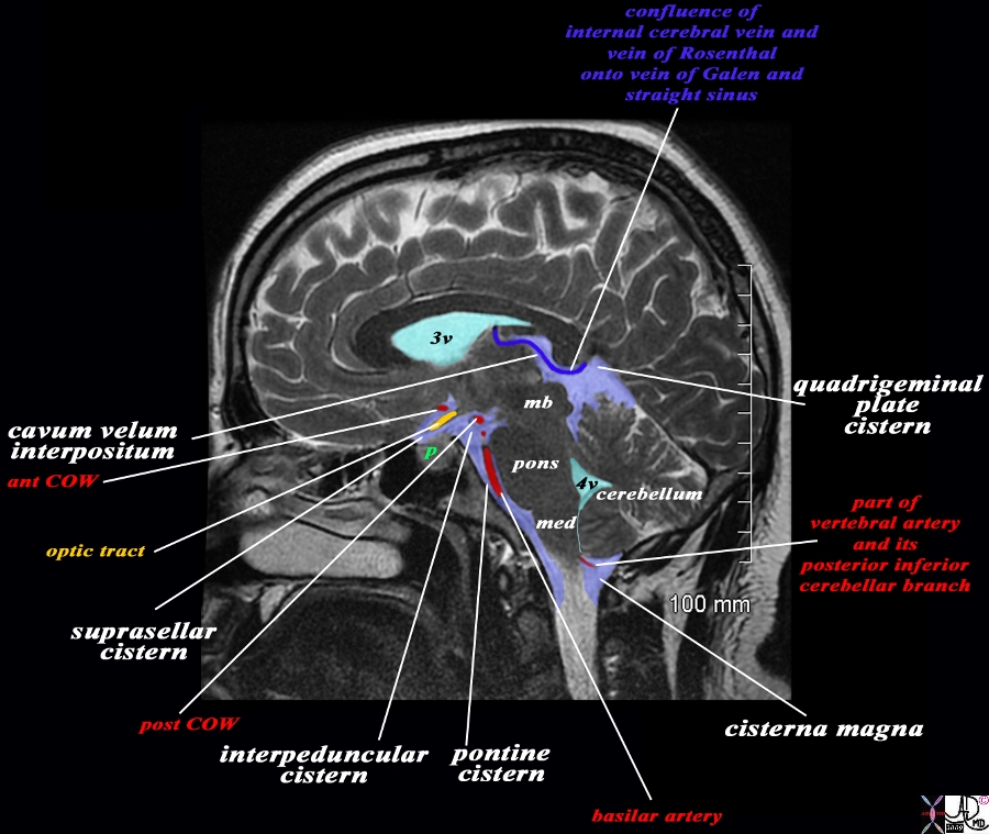
Vessels Associated with the Cisterns |
|
The overlay outlines the basal cisterns in light blue, and the ventricles (teal blue), and a variety of cerebral structures including the midbrain (mb) pons, medulla (med) and cerebellum. A variety of blood vessels (overlaid in blue and red are related to the basal cisterns. The image is T2 weighted sagittal MRI sequence from a patient with a macroadenoma of the pituitary (green p). Starting inferior and anterior we follow the pontine or prepontine cistern associated with the basilar artery, the interpeduncular cistern which is anterior to the peduncles of the midbrain. The suprasellar cistern (above the pituitary – green “p”) is associated with the posterior aspect of the circle of Willis The cavum velum interpositum is associated with the optic tract (yellow) and the anterior vessels of the circle of Willis. Posteriorly the quadrigeminal plate cistern lies behind the colliculi lies anterior to the tentorium where the internal cerebral vein and vein of Rosenthal enter the vein of Galen (blue vein) to enter the straight sinus. Finally the cisterna magna which surrounds the inferior aspect of the medulla and cerebellum merges with the subarachnoid space around the cord where the vertebral artery and posterior inferior cerebellar artery run.
Image Courtesy Ashley Davidoff MD Copyright 2010 All rights reserved 98287b03L03.9s
|
References
Ryan S McNicholas M, and Eustace S. Anatomy for Diagnostic Imaging Second Edition Elsevier Health Sciences