
The Common Vein Copyright 2010
Introduction
The blood supply of the brain is ultimately carried back via the internal jugular veins. The cerebral venous system is a system without valves. The dural sinuses also play a major role in the venous drainage of the brain. As mentioned, in some areas of the dura, the two layers of the dura split and form a venous sinus. They are the superior and inferior sagittal sinuses, the straight sinuses and the two transverse sinuses. All the sinuses, except the inferior sagittal sinus converge at the confluence of sinuses. The inferior sagittal sinus reaches the confluence by way of the straight sinus. Each transverse sinus continues on as a sigmoid sinus, which eventually drains into the internal jugular vein as it exits the jugular foramen. The venous drainage originates from two sources. The deep cerebral veins empty drain the deep cerebral structures and empty into the straight sinus, one of the dural sinuses. The internal cerebral vein is one of the major veins of the deep venous system. The two internal cerebral veins converge to form the great vein of Galen. This vein of Galen then forms the straight dural sinus when it converges with the inferior sagittal sinus. The great vein of Galen is also responsible for draining the basal veins, which receive the drainage of the insula and parts of the basal ganglia. The superficial venous drainage consists of veins that drain into the superior and inferior sagittal sinuses. The drainage then follows posteriorly towards the internal jugular. The dural sinuses also play another important role in maintaining the fluid balance within the calvarium. The dural sinus, especially in the superior sagittal sinus, there is active reabsorption of CSF via arachnoid villi/granulations. These arachnoid villi can be damaged by hemorrhage or infection and lead to increased CSF in a non-obstructive type of hydrocephalus.
The Venous System – Concepts |
|
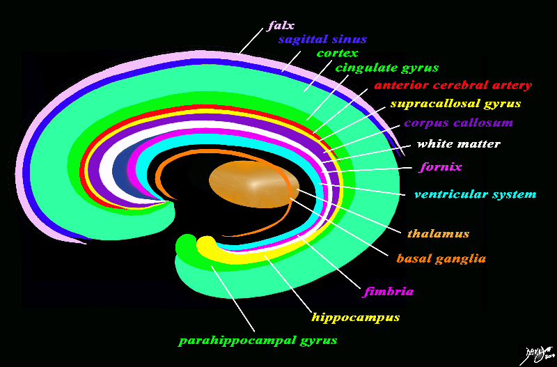
The forebrain has most of its components aligned in a series of inverted c- shaped rings starting from the outer membranes that culminate in the falx (pink), then extending inward smaller inner rings with each intimately connected to the others. The venous system follows this pattern in that the superior sagittal sinus and the inferior sagittal sinus both follow the inverted C concept. In this diagram the the “sagittal sinus” is conceptua;ised in relation to the falx The thalamus (dull orange) appears diagrammatically as the centre of these rings as seen from the sagittal view The outer ring is the falx (pink) followed by the sagittal sinus (blue) cerebral cortex (light green), cingulate gyrus (bright green) superiorly which becomes the parahippocampal gyrus inferiorly. The red ring represents the distribution of the main portion of the anterior cerebral artery. Next is the yellow ring which is the supracollosal gyrus (indusium griseum) superiorly and the hippocampus inferiorly. This is followed by the corpus callosum (purple) which enables the white ring of white matter to connect between hemispheres. The next ring is the thin bright pink ring which represents the fornix superiorly and the fimbria inferiorly. The innermost ring (light blue) represents the lateral horns of the ventricular system. The basal ganglia run just lateral to the lateral ventricles. The navy blue arrow headed structure is the septum pellucidum. Courtesy Ashley Davidoff copyright 2010 all rights reserved 93907d13g03.8s |
Dural Sinuses and Cerebral Veins

Venous Drainage – Lateral Projection |
| 49421 hx 49 F with headaches artery brain cerebral circulation carotid artery internal carotid venous phase superior sagittal sinus vein of Galen vein of Rosenthal transverse sinus normal anatomy lateral projection arteriogram arteriography angiogram angiography Davidoff MD 49400, 49401 49402, 49403, 49409, 49410, 49411,49416, 49417, 49418, 49419, 49420, 49421, 49422, 49424 |

Venous Drainage – A-P Projection |
| 49424 hx 49 F with headaches artery brain cerebral circulation carotid artery superior sagital sinus vein of Galen vein of Rosenthal transverse sinus sigmoid sinus internal carotid injection venous phase normal anatomy arteriogram arteriography angiogram angiography Davidoff MD 49400, 49401 49402, 49403, 49409, 49410, 49411,49416, 49417, 49418, 49419, 49420, 49421, 49422, 49424 |

Intracranial Veins -a source of headaches |
| This venous phase of the angiogram in the antero-posterior projection is normal. Venous evaluation is not performed in the workup of headches but the intracranial veins are resposible for some headaches. The eact pathogeneseisis remains controversial and is a source of ongoing research.
49424c01 brain cerebral veins arachnoid granulations superior sagittal sinus thalamostriate veins transverse sinus sigmoid sinus basal vein of Rosenthal jugular bulb jugular vein venous phase of A-P angiogram normal anatomy angiography Courtesy Ashley Davidoff MD |
|
MRV – Dural Sinuses and Veins |
|
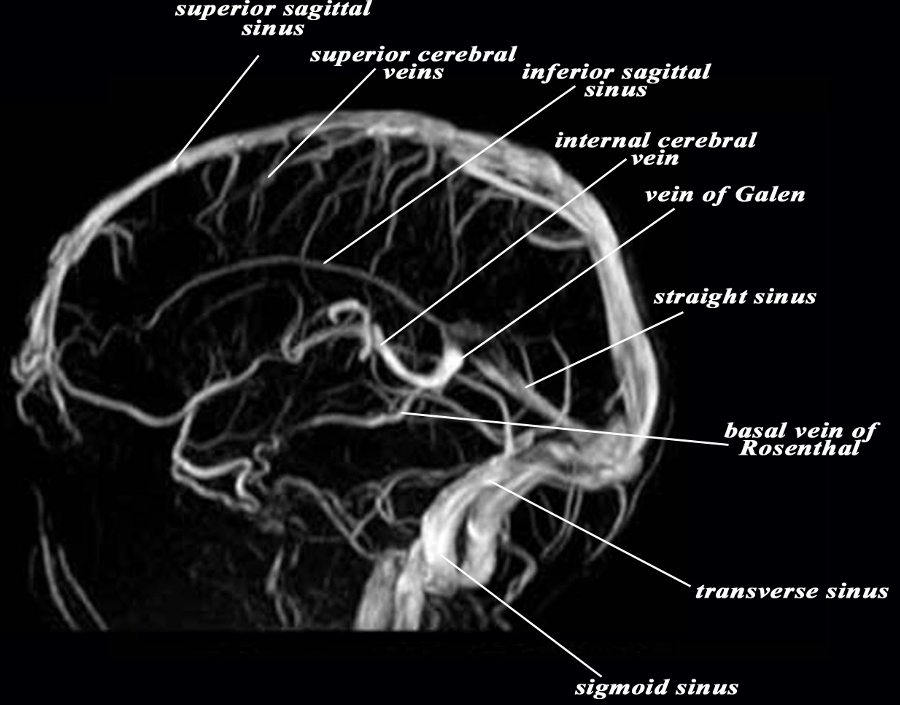
The MRV shows a venogram in the lateral projection demonstrating the dural sinuses and some of the cerebral veins. The superior sagittal sinus runs on the superior aspect of the falx, curves around the posterior parietal region and joins the straight sinus, and transverse sinus to enter the torcular herophili in occipital region. It receives between 10-15 superior cerebral veins. The inferior sagittal sinus is a much smaller vessel and drains into the straight sinus. The sigmoid sinus drains into the internal jugular vein (not shown) The vein of Galen receives the internal cerebral vein and the basal vein of Rosenthal. Image Courtesy Philips Medical Systems 92393.9L |
Venous Drainage Via the Subarachnoid Space |
|
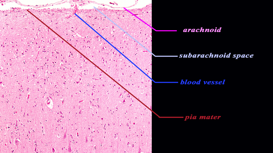
The histological section of the cortex at 10X magnification reveals two of the three meninges of the brain. The inner layer (maroon) is the pia mater and it is in intimate contact with the brain and faithfully follows the sulci and gyri. At this magnification it is barely visible since it is very thin. The second layer is the arachnoid (pink) which is a slightly thicker membrane and follows the pia in a general fashion but does not extend into the sulci. It is intimately attached to the inner layer of the dura The space between the pia and the arachnoid is the subarachnoid space. The blood vessel seen entering the space is likely a small vein. Courtesy of Thomas W.Smith, MD; Department of Pathology, University of Massachusetts Medical School 97377b04.9. |
Diseases Applied Biology
|
Venous Angioma Around the Cerebellum |
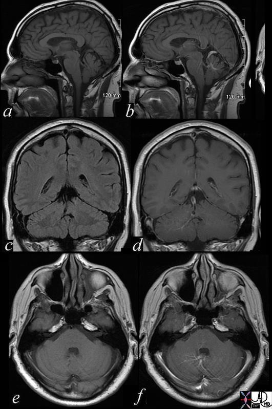
| 71730c02 brain cerebellum vermis vein enlarged venous angioma old bleed superior left vermis – cerebellar venous malformation vascular malformation a = T1 pre gadolinium b = T1 post contrast c = T1 pre gadolinium d = T1 post contrast e = T1 pre gadolinium f = T1 post contrast MRI Davidoff MD |
Acute Falcine and Frontal Subdural Hemorhage |
|
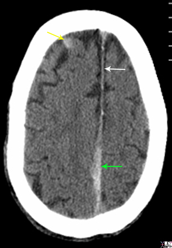
The non enhanced CTscan is from a 80 year old male on coumadin who sustained trauma to the head. There are two acute subdural hemorrhages. The first is a falcine subdural (green arrow) that has resulted in thickening irregularity and increased density of the falx near the vertex. This is contrasted to the normal falx anteriorly (white arrow) The second acute subdural is in the right frontal region (yellow arrow). Courtesy Ashley Davidoff MD Copyright 2010 89283.8sL |