Hindbrain – Transverse or Axial Projection – Concepts
The Common Vein Copyright 2010
Introduction
The Pons
The Pons |
|
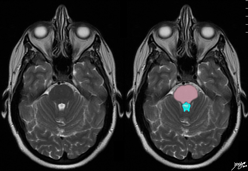
The conceptual framework of the pons is overlaid on a T2 weighted MRI through the most characetristic axial image of the pons. This location is directly anterior to the widet portion of the aqueduct of Sylvius posteriorly (blue) that assumes the shape of a puffy royal crown . Courtesy Ashley Davidoff MD copyright 2010 all rights reserved 94084c01.81s |
The Pons The 4th Ventricle |
|
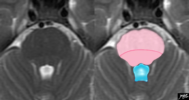
TheT2 weighted MRI is a magnified view of the pons recognized commonly in the axial projection by its relationship to the 4th ventricle posteriorly which has a characteristic shape looking like a puffed up crown (blue) The pons consists of an anterior portion called the ventral pons (light pink) and the dorsal portion called the tegmentum ((darker pink). The line of distinction is vague and the dark pink line is drawn in for clarity. Courteys Ashley Davidoff MD copyright 2010 all rights reserved 94084.3kdc.82sd01 |

The Pontiff’s Hat that Sits on the Pons |
|
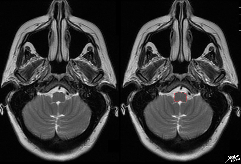
The 4th ventricvle in the axial plane looks like the pontiff’s hat which is called the mitre A pmnemonic you may want to use to remind you of the structure that is posteriorly related to the 4th ventricle may be “The pontiff’s hat that sits on the pons” Copyright Ashley Davidoff MD Copyright 2010 all rights reserved 82407b14d05.8s |
The Medulla
The Medulla |
|
The medulla in the axial plane on this T 2 weighted MRI is almost rectangular in shape but is characterized by the two sets of bulges anteriorly. The medulla is divided into the anterior ventral portion portion and the posterior tegmentum. The anterior portion consists of the medial pyramidal tracts and the lateral olives . It is these two structures that account for the symmetrically positioned anterior bulges. Courtesy Ashley DAvidoff MD copyright 2010 all rights reserved 94086b01.3kc.8s |
Conceptual Components of the Medulla |
|
The medulla in the axial plane on this T 2 weighted MRI is almost rectangular in shape but is characterized by the two sets of bulges anteriorly. The medulla is divided into the anterior ventral portion portion and the posterior tegmentum. The anterior portion consists of the medial pyramidal tracts and the lateral olives Courtesy Ashley DAvidoff MD copyright 2010 all rights reserved 94086b01.3kc01.8s |
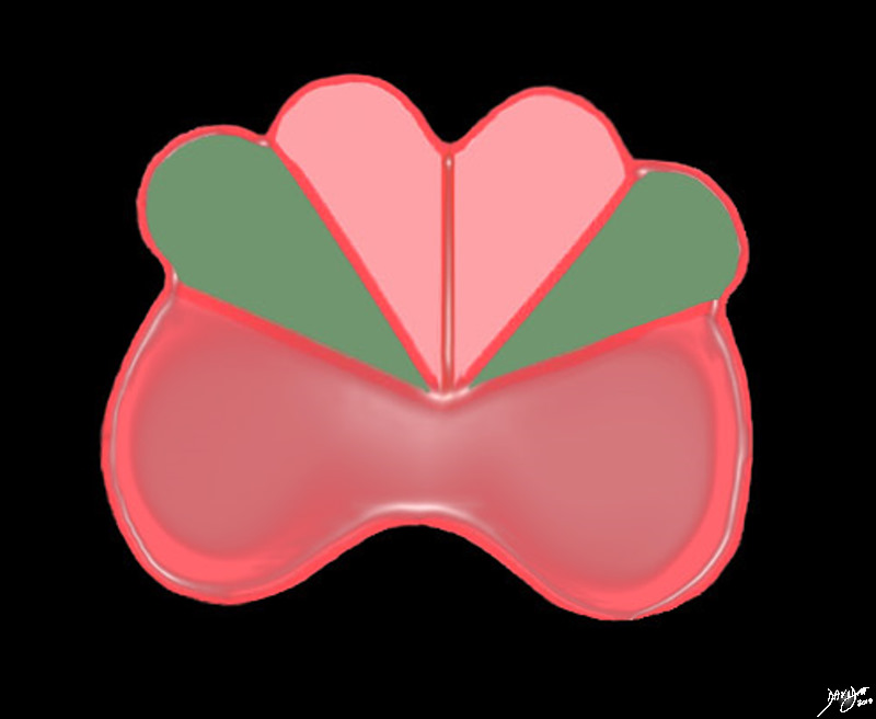
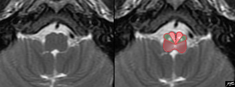
The Medulla 4 Ice Cream Cones in a Row |
|
The medulla at its classic location has some subtle shape diferences when compared to the midbrain and pons mostly accentuated by the olives (green)which are the second set of ice cream shaped wedges . The medial set of ice cream cones that are light pink are the medullary pyramids Posteriorly – the salmon pink bilobed section is called the tegmentum Courtesy Ashley DAvidoff MD copyright 2010 all rights reserved 94086b01.3kb02bb04.8s |
|
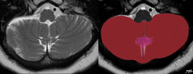
A Heart or a Top |
|
The transverse Tt2 weighted MRI through the main portion of the cerebellum in the posterior cranial fossa reflects a heart shaped structure created with some artistic licence. The cerebellum consists of two cerebellar hemispheres and a central worm like vermis Courtesy Ashley Davidoff MD copyright 2010 all rights reserved 49037.3kb01c.8s |
|
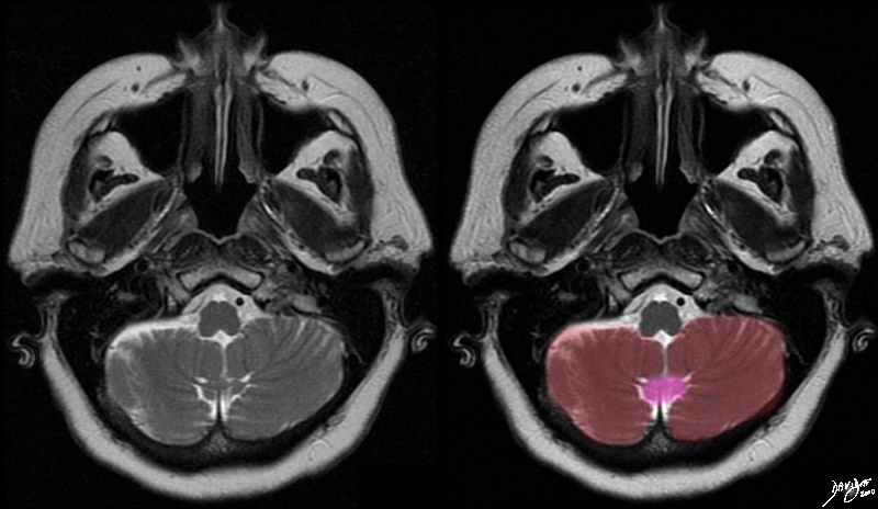
The Hemispheres and the Vermis |
|
The T2 weighted MRI shows the 2 major components of the cerebellum – the central vermis (pink), and the two cerebellar hemispheres. The cerebellum consists of two cerebellar hemispheres and a central worm like vermis Courtesy Ashley Davidoff MD copyright 2010 all rights reserved 49037c.8s |
|
Winking Tabby Cat |
|
The Winking Cat The MRI of the posterior fossa and cerebellum was rendered and reminded the author of a winking tabby cat Davidoff Art Copyright 2010 all rights reserved 49037.3kb03.81s |