
The Common Vein Copyright 2010
Definition
The occipital lobe is part of the forebrain and is located at the rear of the skull, below the parietal lobe and it is the smallest of the four lobes.
Structure The occipital lobe is all the portion of the hemisphere located posterior to the parieto-occipital sulcus. It rests on the tentorium cerebelli. Two fissures (transverse and lateral occipital sulci) separate another 3 convolutions (superior, inferior and lingual gyri). In addition, the lunate sulcus lies vertically in front of the occipital pole. Its lips separate striate from peristriate areas; the parastriate area is in this sulcus between the striate areas.
Function The occipital lobes are the center of our visual perception system. The occipital cortex contains the primary visual cortex. Each half of this cortex receives input from the nasal half of the contralateral visual field and the temporal half of the ipsilateral visual field. It allows for recognition and outlines of images. The cortex devoted to visual analysis extends to the parietal and temporal lobes.
The peristriate region in the occipital lobe is involved in visual spatial processing, discrimination of movement and color discrimination.
Disease The occipital lobes are particularly vulnerable to injury due to their location at the back of the brain, although any significant trauma to the brain could produce subtle changes in our visual-perceptual system, which generates defects and visual field scotomas.
Damage to one side of the occipital lobe can cause homonymous vision loss with the same visual field from both eyes being cut out. Occipital lobe disorders can cause visual hallucinations and delusions. Visual hallucinations (visual images without external stimuli) can be caused by lesions in the occipital region. Visual illusions (distorted perceptions) can take the form of objects that look larger or smaller than they really are, without color objects or objects having abnormal coloring. Dysfunction can result in vision loss or hallucinations. Less severe dysfunction results in the inability to recognize or accurately perceive objects.
Treatment is often surgical for the removal of lesions.
The Occipital Lobe (lime green) Part of the Forebrain (Prosencephalon) Member of the Cerebrum (Telencephalon) Member of the Cerebral Hemispheres
|
|
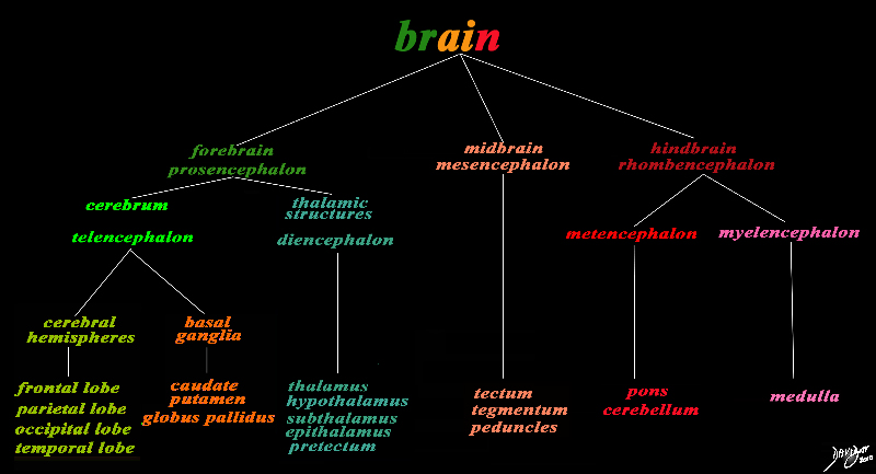
The basic and simplest classification of the brain into forebrain midbrain and hindbrain is shown in this diagram and advanced to a more complex tree using the embryological and evolutionary terminologies. The forebrain consists of the cerebrum also called the prosencephalon, which contains the more advanced form of the brain and the thalamic structures which contain more basic structures. The cerebrum (telencephalon) itself consists of two cerebral hemispheres and paired basal ganglial structures. Each cerebral hemisphere will have gray and white matter distributed in the frontal parietal temporal and occipital lobe, with the basal ganglia being part of the gray matter deep in the cerebral hemispheres. The most important thalamic structures arising from the diencephalons include the thalamus itself and the hypothalamus. The midbrain (mesencepaholon) consists of the tectum tegmentum and cerebral peduncles. The hindbrain has two major branch points based on the evolutionary development. The pons and cerebellum(part of the metencephalon) are grouped and the medulla (part of the myelencephalon is the second branch. Courtesy Ashley Davidoff MD Copyright 2010 All rights reserved 97686.8s |
|
Conceptual Framework of the Brain |
|
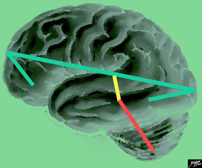
The vector of the forebrain (green) is folded at its anterior aspect as a projection of the frontal lobe and the posterior fold forms the temporal lobe. The midbrain (yellow has its axis just tipped slightly anteriorly and the hindbrain (salmon red) is tipped more posteriorly Courtesy Ashley Davidoff MD copyright 2010 all rights reserved |
The Central Sulcus is the Border between the Frontal Lobe and the Parietal Lobe |
|
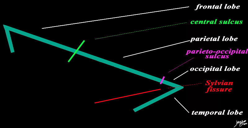
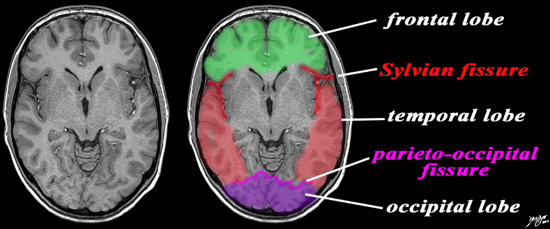
The forebrain is divided into the frontal lobe anetriorly, separated by the central sulcus (bright green), the parietal lobe separated from the occipital lobe by the parieto-occipital fissure (pink), and the temporal lobe separated from the frontal and occip[ital lobe by the Sylvian fissure (lateral sulcus) (red). The distinction between the occipital and temporal lobe is more of a functional division than a structural division Courtesy Ashley Davidoff MD copyright 2010 93887b03b05.8s |
|
83029d13.8s |
|
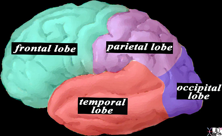
This is a diagram looking at the forebrain from the side showing the anteriorly placed frontal lobe, inferiorly placed temporal lobe posteriorly placed occipital lobe and superiorly placed occipital lobe. In this view the size of the frontal lobe dominates and in fact it is the largest portion of the forebrain. Courtesy Ashley DAvidoff MD copyright 2010 all rights reserved 83029d13.8s |
The Occipital Lobe defined by the Parieto-Occipital Sulcus |
|
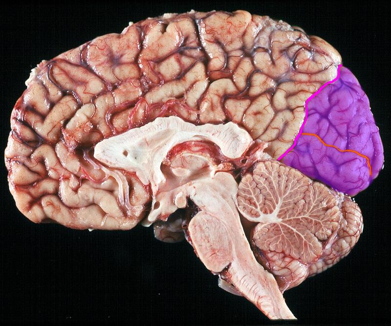
The sagittal view of the anatomical specimen of the brain shows the parieto-occipital fissure (pink) (aka sulcus), that separates the parietal lobe anteriorly and the occipital lobe posteriorly (purple). The calcarine fissure (orange) separates the cuneus above from the lingual gyrus inferiorly. Image Courtesy of Thomas W.Smith, MD; Department of Pathology, University of Massachusetts Medical School. 97805bd04 |
Occipital Lobe defined by the Parieto-occipital Fissure |
|
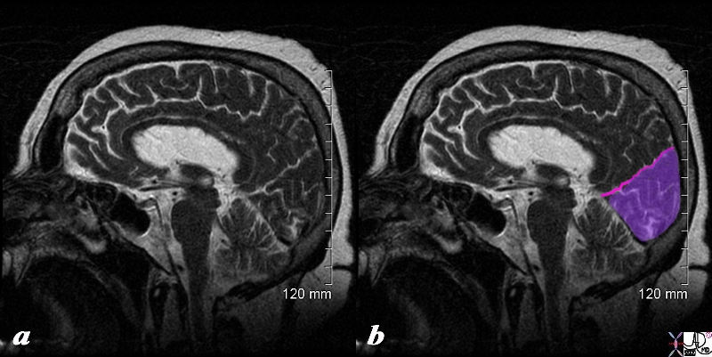
The sagittal view of the brain using a T2 weighted sequence shows the parieto-occipital fissure (pink) (aka sulcus), that separates the parietal lobe anteriorly and the occipital lobe (purple) posteriorly. Courtesy Ashley Davidoff copyright 2010 all rights reserved 71430cd01b.8s |
Occip[itoparietal Fissure Calcarine Fissure and Occipital Gyri |
|
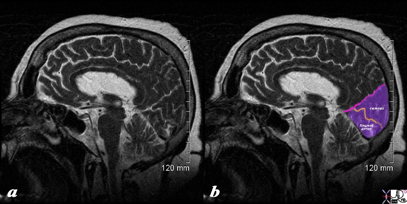
The sagittal view of the brain using a T2 weighted sequence shows the parieto-occipital fissure (pink) (aka sulcus), that separates the parietal lobe anteriorly and the occipital lobe (purple) posteriorly. The calcarine fissure (orange) separates the cuneus above from the lingual gyrus inferiorly. Courtesy Ashley Davidoff copyright 2010 all rights reserved 71430cd01b02.8s |
|
Axial Framework of the Forebrain |
|
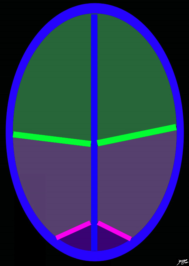
The diagram reflects the interhemispheric fissure as depicted in the axial plane dividing the brain into two halves. The bright green line represents the central sulcus which separates the the frontal lobes (green) from the parietal lobes (light purple. The parito-occipital fissure (pink) separates the parietal lobes from the occipital lobes. By convention anterior is on the top and posterior on the bottom, while the patients right is to our left and vice versa for the patients left Copyright 2010 Courtesy Ashley Davidoff MD 93914.85sb |
|
Axial Projection MRI |
|
The axial image is intended to demonstrate ttwo important fissures; the Sylvian fissure (in red) the parieto-occipital fissure (pink) in order to demonstrate the border between the frontal and temporal lobe in the axial plane and the junction of the temporal and occipital lobe MRI SENSE Courtesy Philips medicaL systems rendred by Davidoff art 92142c06b01 label.8s |
Applied Anatomy Diseases
Ischemia
Subtle Occipital Lobe Loss of Gray White Differentiation |
|
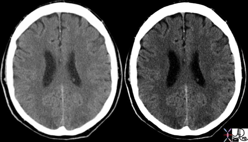
This hypertensive patient presents with visual disturbance and the axial CT scan shows subtle changes in the occipital lobe characterised by vague low density regions. There is loss of gray-white matter differentiation consistent with acute ischemia. These findings are consistent with acute hypertensive encephalopathy Courtesyt Ashley Davidoff MD 73422 |
|
Acute Multicentric Non Hemorrhagic Infarcts Consistent with Embolic Disease |
|
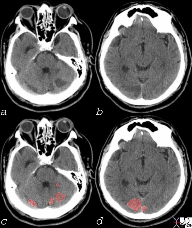
Multicentric Infarcts in the Cerebellum and Occipital Lobes Multicentric low density regions in both posterior cerebellar hemispheres and the occipital lobes bilaterally seen in (a and b) correspondingly overlaid in light red in c and d, are consistent with non hemorrhagic infarcts. Embolic disease is most likely Courtesy Ashley Davidoff MD 74923c01 |
FLAIR Hyperintensity in the Occipital Lobes |
|
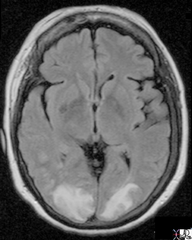
This hypertensive patient presents with visual disturbance and the MRI using a FLAIR sequence shows obvious FLAIR hyperintensity in the occipital lobes and extending into the parietal lobes. These findings are consistent with acute hypertensive encephalopathy and ischemia of the occipital lobes characteristic of acute hypertensive encephalopathy Courtesy Ashley Davidoff MD Copyright 2010 All rights reserved 73422 |