
The Common Vein Copyright 2010
Definition
The forebrain consists of the
cerebral hemispheres (cerebrum)
the basal ganglia and the
thalamus and thalamic structures including the hypothalamus.
It is characterized by being the largest part of the brain. It is that part of the brain that lies in the most anterior and superior part of the cranial cavity, and
Embryologically it consists of the structures of the;
telencephalon (aka cerebrum) which consists of the two cerebral hemispheres and basal ganglia
diencephalon are the thalamus and other thalamic structures – (hypothlamus prethalamus, subthalamus, and epithalamus)
The Forebrain in Context |
|
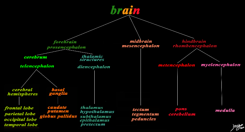
The basic and simplest classification of the brain into forebrain midbrain and hindbrain is shown in this diagram and advanced to a more complex tree using the embryological and evolutionary terminologies. The forebrain consists of the cerebrum also called the prosencephalon, which contains the more advanced form of the brain and the thalamic structures which contain more basic structures. The cerebrum (telencephalon) itself consists of two cerebral hemispheres and paired basal ganglial structures. Each cerebral hemisphere will have gray and white matter distributed in the frontal parietal temporal and occipital lobe, with the basal ganglia being part of the gray matter deep in the cerebral hemispheres. The most important thalamic structures arising from the diencephalons include the thalamus itself and the hypothalamus. The midbrain (mesencepaholon) consists of the tectum tegmentum and cerebral peduncles. The hindbrain has two major branch points based on the evolutionary development. The pons and cerebellum(part of the metencephalon) are grouped and the medulla (part of the myelencephalon is the second branch. Courtesy Ashley Davidoff MD Copyright 2010 All rights reserved 97686.8s |
Converting the Table into a Diagram
Basic Components of the Forebrain |
|
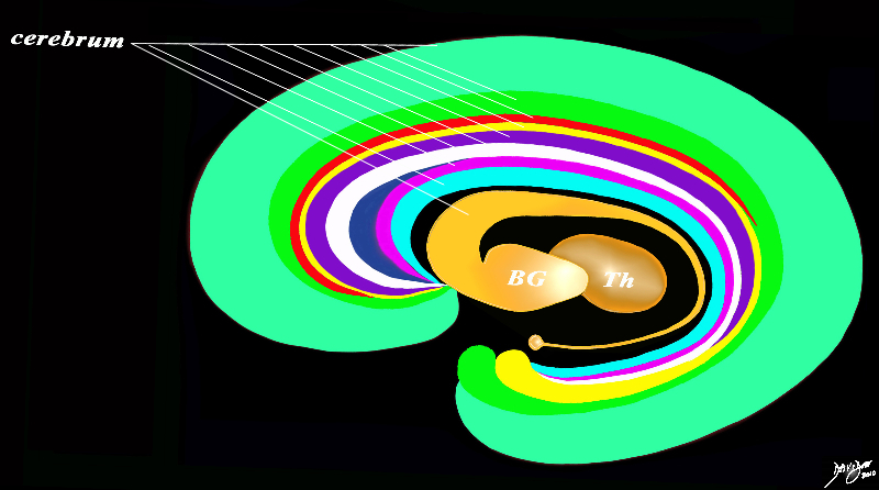
The diagram reflects the two basic parts of the forebrain including the; cerebrum (2 cerebral hemispheres – telencephalon) and the thalamic structures (Th – dark orange – diencephalon). The cerebrum (telencephalon) consists of the cerebral hemispheres and the group of basal ganglia structures Conceptually the cerebrum and cerebral hemispheres (telencephalon) consist of two large groups: a) The outer coat; (of many colors) including the frontal lobe, parietal lobe, temporal lobe, and occipital lobe in which are housed the series of inverted “C-rings” such as the fornix, white matter, corpus callosum, cingulate cortex, hippocampus and parahippocampal gyrus. b) The inner group of basal ganglia (light orange) consisting in general of the caudate nucleus, putamen and globus pallidus. The thalamus and the thalamic structures complete the components of the forebrain. The thalamic structures (diencephalon) include the thalamus hypothalamus, subthalamus, epithalamus and subtectum. Courtesy Ashley Davidoff MD Copyright 2010 All rights reserved 93907d13b05b02h.81s |
Sagittal View of the Anatomy |
|
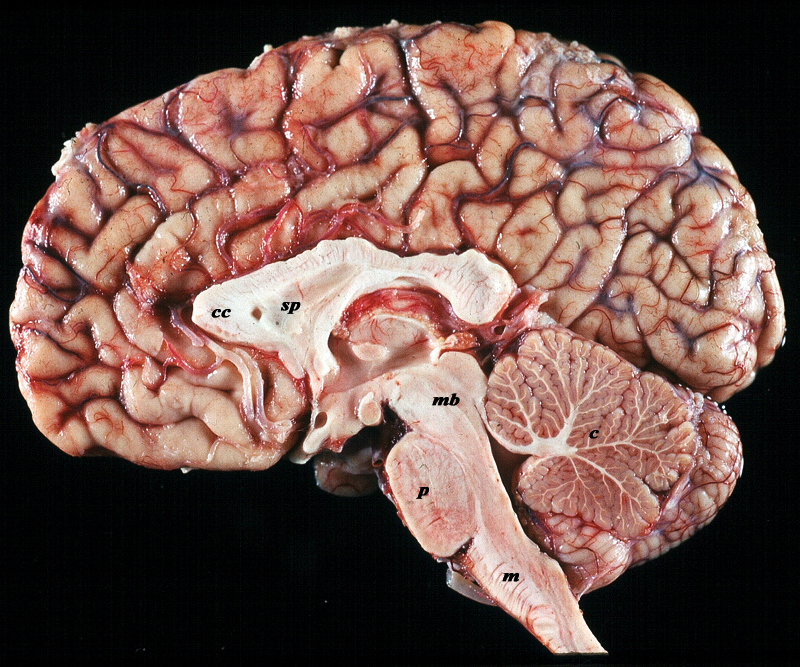
The midsagittal section view of brain reveals the distinctive shape position and character of the midline structures of the brain. The distinction between the character of the cerebral cortex which has a creamy color and the white matter exemplified by the corpus callosum (c) and septum pellucidum (sp) which are white, and the midbrain (mb) pons (p) and medulla (m) which are off white as opposed to the color of the cerebellum (c) which is light salmon pink is well demonstrated. The relative sizes of the forebrain, midbrain and hindbrain and their components are well appreciated in this section. Image Courtesy of Thomas W.Smith, MD; Department of Pathology, University of Massachusetts Medical School. 97805b02 |
Functionally the forebrain is the part of the brain that is essential to our survival in day to day activity and enables all the supratentorial processes that allow us to flourish as a species. The forebrain in general controls cognitive function which represents the aspect that makes us human including the processes of knowing, thinking, learning, and judging. It also is central to emotional, sensory and motor function. Basic bodily functions of eating, sleeping, temperature regulation and reproductive functions are also controlled by the forebrain.
Common diseases that affect the forebrain include strokes, tumors, degenerative disorders, infections and inflammatory disorders.
Diagnosis of disease manifests clinically with aberrance in cognitive, motor, and sensory function. Imaging is commonly accomplished by MRI and CT scanning
Treatment includes both medical and surgical disciplines.
Structurally it consists of the cerebral hemispheres, (frontal lobe, parietal lobe temporal lobe and occipital lobe), corpus callosum, basal ganglia, limbic system, thalamus and the hypothalamus.
The Forebrain – Largest Part of the Brain |
|
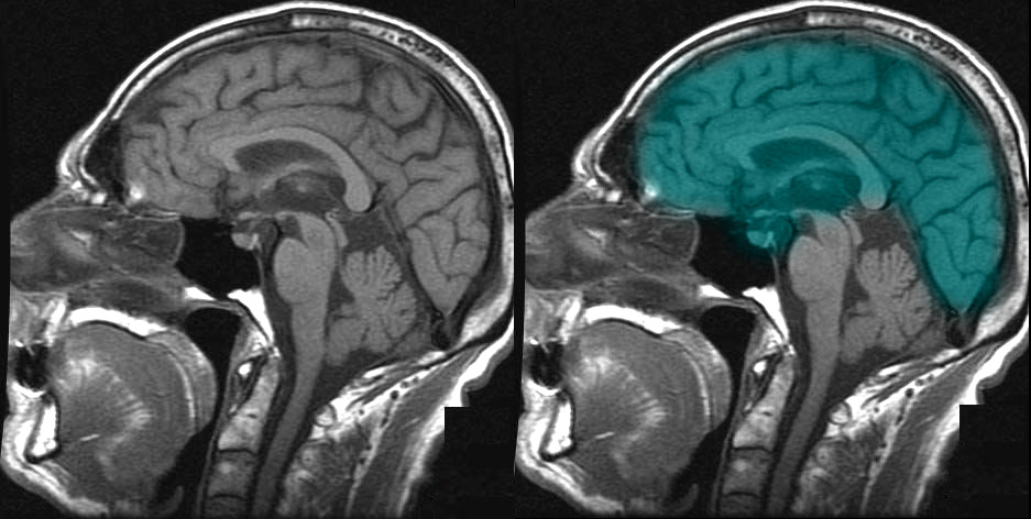
The T1 weighted MRI taken in sagittal projection reflects the forebrain (torquoise). It is the largest and most superior part of the brain and occupies the anterior cranial fossa. Courtesy Ashley Davidoff MD copyright 2010 all rights reserved 49079b01b02b01 |
|
Shapes on the Top |
|
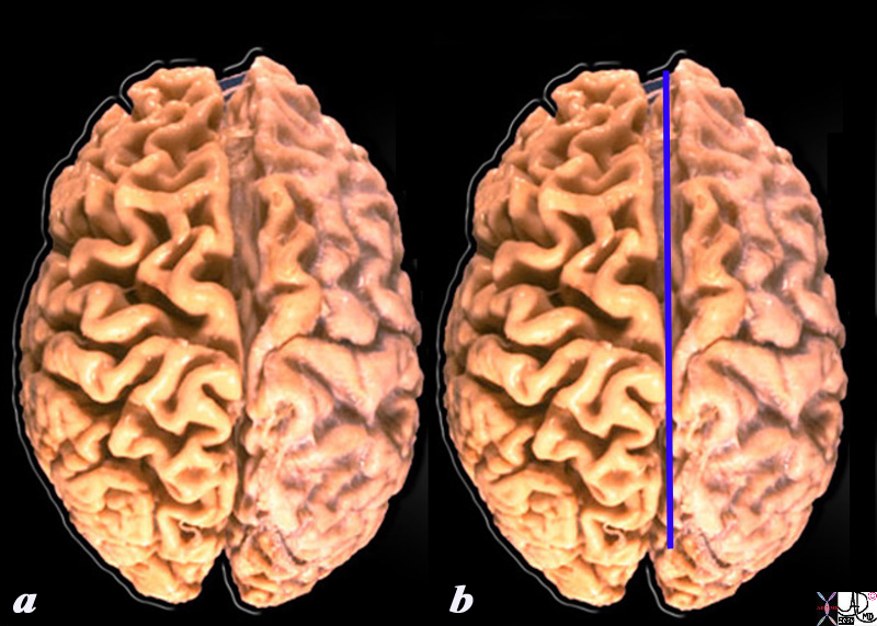
When the external surface is explored from above a deep midline fissure (blue line) divides the oval of the brain into two hemispheres. The deep cleft is called the interhemishpheric fissure In addition, the serpiginous folds and crevices that characterize the outside surface of the brain start to become apparrent as one explores the shape of the brain in more depth. It becomes obvious that it is more than a mere oval. Source unknown Image modified by Davidoff 52981b05d05 |
|
T2 weighted MRI The Interhemispheric Fissure |
|
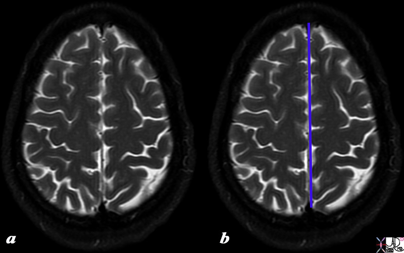
The T2 weighted MR taken a few cms from the vertex demonstrates the overall ovoid shape of the brain, the interhemishperic fissure, the sulci filled with fluid (white) enabling visualization of the gyri (black) Courtesy Ashley DAvidoff MD copyright 2010 all rights reserved 90720c01.8s |
|
Coronal View The Interhemispheric Fissure |
|
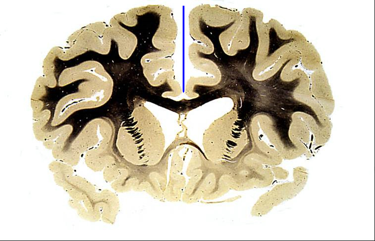
The coronal section of a normal anatomic section of the brain shows the interhemispheric fissure (blue line) and in particular the depth to which it extends into the brain. Courtesy Elisa Flower MD Department of Radiology Boston Medical Center and Courtesy of Department Anatomy Boston University School of Medicine 97104b02 |
The Lobes |
|
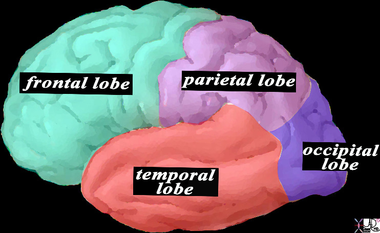
This is a diagram looking at the forebrain from the side showing the anteriorly placed frontal lobe, inferiorly placed temporal lobe posteriorly placed occipital lobe and superiorly placed occipital lobe. In this view the size of the frontal lobe dominates and in fact it is the largest portion of the forebrain. Courtesy Ashley DAvidoff MD copyright 2010 all rights reserved 83029d13.8s |
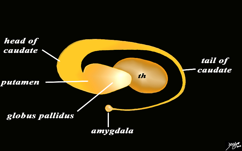
The Basal Ganglia |
|
The basal ganglia lie both in the forebrain and in the midbrain. In the forebrain they are distributed in paraventricular fashion almost in an inverted C fashion. In the conceptual diagrams we have colored the inverted “C” that abuts the ventricular system orange, and this includes the thalamus and the basal ganglia. The basal ganglia that lie in the forebrain include the globus pallidus, putamen, head of the caudate nucleus, tail of the caudate nucleus and the amygdala. The amygdala appears to be part of the limbic system and the basal ganglia. Courtesy Ashley Davidoff MD Copyright 2010 All rights reserved 93907d13b05dL3.8s |