
Centrum Semiovale
Copyright 2010
Definition
Centrum Semiovale |
|
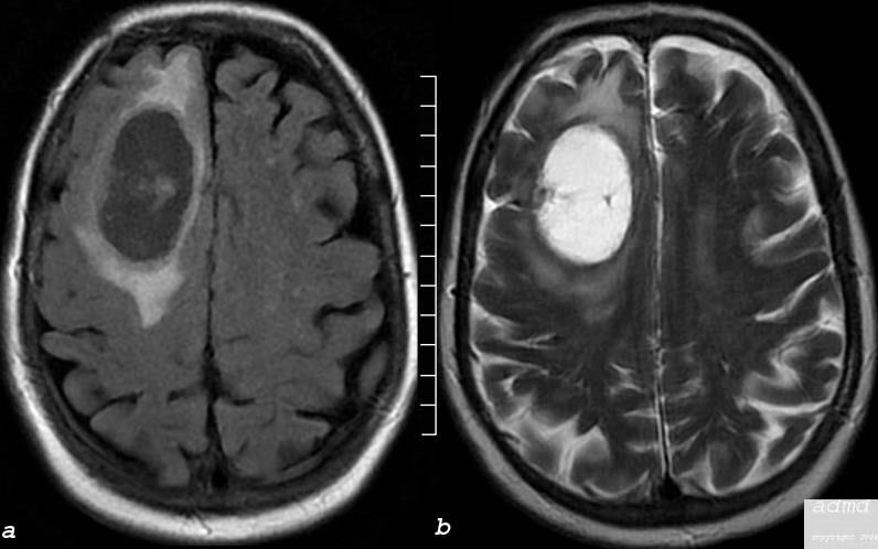
The MRI performed in the axial plane shows a large mass in the right frontal region and the anterior half of the centrum semiovale. Image (a) is an axial image through the vertex of the brain using FLAIR technique showing mostly a cystic type lesion with some internal thick sepatation with surrounding vasogenic edema. Minimal mass effect on the midline is perceived. Image (b) is an axial T2 weighted sequence showing a cystic matrix and surrounding edema The lesion was shown to be a cavitating squamous cell metastasis brain frontal lobe mass cystic moderate surrounding edema consistent with squamous cell carcinoma metastasis edema involves the anterior half of the right centrum semiovale Courtesy Ashley Davidoff MD Copyright 2010 75408c03 |